. 2017 Feb;113(2):234-246.
doi: 10.1093/cvr/cvw249. Epub 2017 Jan 14.

Mitochondrial redox plays a critical role in the paradoxical effects of NAPDH oxidase-derived ROS on coronary endothelium

Affiliations

Mitochondrial redox plays a critical role in the paradoxical effects of NAPDH oxidase-derived ROS on coronary endothelium

Ehtesham Shafique et al. Cardiovasc Res. 2017 Feb.

Abstract

Aims: There are conflicting reports on the role of reactive oxygen species (ROS) i.e. beneficial vs. harmful, in vascular endothelium. Here, we aim to examine whether duration of exposure to ROS and/or subcellular ROS levels are responsible for the apparently paradoxical effects of oxidants on endothelium.

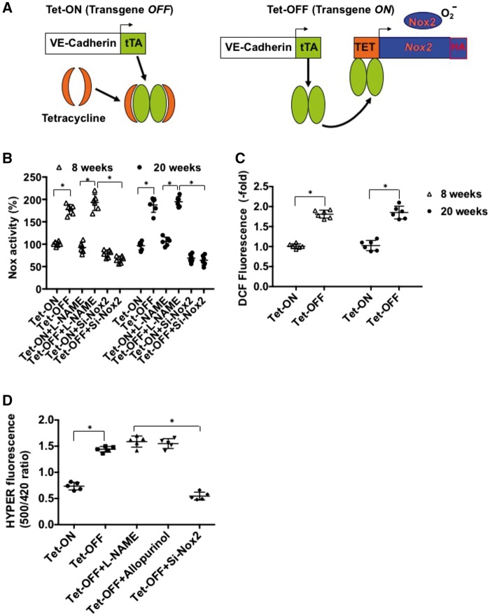
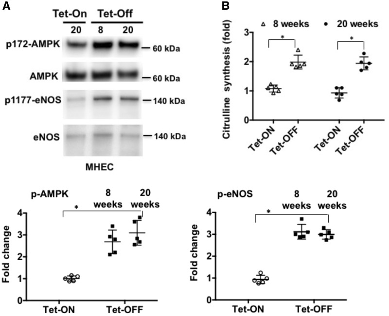
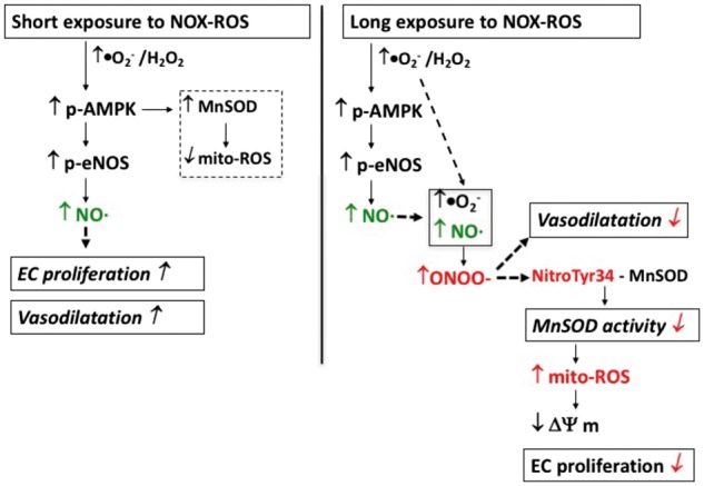
Methods and results: We have recently generated binary (Tet-ON/OFF) conditional transgenic mice (Tet-Nox2:VE-Cad-tTA) that can induce 1.8 ± 0.42-fold increase in NADPH oxidase (NOX)-derived ROS specifically in vascular endothelium upon withdrawal of tetracycline from the drinking water. Animals were divided in two groups: one exposed to high endogenous ROS levels for 8 weeks (short-term) and the other for 20 weeks (long-term). Using endothelial cells (EC) isolated from mouse hearts (MHEC), we demonstrate that both short-term and long-term increase in NOX-ROS induced AMPK-mediated activation of eNOS. Interestingly, although endothelium-dependent nitric oxide (NO)-mediated coronary vasodilation was significantly increased after short-term increase in NOX-ROS, coronary vasodilation was drastically reduced after long-term increase in ROS. We also show that short-term ROS increase induced proliferation in EC and angiogenic sprouting in the aorta. In contrast, long-term increase in cytosolic ROS resulted in nitrotyrosine-mediated inactivation of mitochondrial (mito) antioxidant MnSOD, increase in mito-ROS, loss of mitochondrial membrane potential (Δψm), decreased EC proliferation and angiogenesis.

Conclusion: The findings suggest that NOX-derived ROS results in increased mito-ROS. Whereas short-term increase in mito-ROS was counteracted by MnSOD, long-term increase in ROS resulted in nitrotyrosine-mediated inactivation of MnSOD, leading to unchecked increase in mito-ROS and loss of Δψm followed by inhibition of endothelial function and proliferation.

Keywords: Endothelium • Signal transduction • Nitric oxide • Reactive oxygen species • NADPH oxidase.


Figures

Figure 1
Figure 1
Endothelium-specific overexpression of Nox2 and maintenance of increased ROS generation in coronary ECs for 8 (short-term)–20 (long-term) weeks. (A) Schematic presentation of conditional binary transgenic mice (NVF) generation. Tet-ON, tetracycline was added to the drinking water to suppress expression of the Tet-Nox2 transgene (left panel); Tet-OFF, withdrawal of tetracycline to induce the transgene (right panel). (B) NADPH oxidase activity in ECs isolated from mouse hearts (MHEC) was measured using low-concentration lucigenin (5 μM) and 100 µM NADPH. Tet-OFF animals were without tetracycline for eight or 20 weeks as indicated. MHEC were transfected with siRNA against Nox2 or pre-incubated with NOS-inhibitor l-NAME (500 μM), where indicated. (C) H2DCF-DA fluorescence assay as a measure of total intracellular ROS in MHECs. (D) H2O2-sensitive Hyper-2 probe was used to measure intracellular ROS in MHECs as indicated. Where indicated, MHEC Tet-OFF for 20 weeks were pre-incubated with L-NAME (NO synthase inhibitor; 500 µM), allopurinol (xanthine oxidase inhibitor; 100 μM) or transfeted with si-Nox2 for specificity of the probe. Tet-ON MHECs were grown in medium containing tetracycline 2 μg/mL. All assays were carried out in triplicate. Scatter plot data shown are of triplicate experiments using independent replicates of n = 5 or more batches of ECs as indicated. *P < 0.05 (N ≥ 5/time point/group).
Figure 2
Figure 2
Both short (8 weeks)- and long (20 weeks)-term increase in endogenous ROS levels activate AMPK and eNOS in ECs. (A) Western blots (WB) of MHEC protein lysates from NVF Tet-ON (20 weeks) and Tet-OFF (8 and 20 weeks) mice were performed as indicated. WB was carried out using anti-phospho 172-AMPK (p-AMPK), anti-p-eNOS (ser1179) and anti-eNOS and anti-AMPK antibodies. Tet-ON MHECs were grown in culture medium containing tetracycline 2µg/mL. Lower panels demonstrate quantitative analyses of p-AMPK and p-eNOS activation in n = 5 independent batches of isolated MHECs performed in triplicates (n= 3 hearts pooled from thre animals per batch of MHEC isolation) and NIH Image J. Densitometry data of p-eNOS and p-AMPK were normalized against total eNOS and total AMPK levels, respectively. *P < 0.05. (B) Nitric oxide (NO) synthesis, as measured using l-NAME (500 µM)-inhibitable citrulline formation assay as described in the Methods section, in 8-week and 20-week Tet-OFF MHEC. Experiments were performed in triplicates using independent batches of MHECs (n = 5) per group and per time points. *P < 0.05.
Figure 3
Figure 3
Short-term increase in endogenous Nox-ROS improves coronary vasodilatation but long-term increase in Nox-ROS does not. (A) Endothelium-dependent dilation of coronary arterioles from Tet-ON (n = 8 animals per group per time points) and Tet-OFF (for 8 weeks) (n = 8 animals per group per time points) NVF mice in response to VEGF. (B) Endothelium-dependent dilation of coronary arterioles from Tet-ON (n = 8) and Tet-OFF (for 8 weeks) (n = 8) NVF mice in response to acetylcholine (Ach). (C) Endothelium-dependent dilation of coronary arterioles from Tet-ON (n = 6) and Tet-OFF (for 20 weeks) (n = 6) NVF mice in response to VEGF. (D) Endothelium-dependent dilation of coronary arterioles from Tet-ON (n = 6) and Tet-OFF (for 20 weeks) (n = 6) NVF mice in response to acetylcholine (Ach). All coronary vessels were pre-constricted ex vivo using U4669 prior to the addition of VEGF or Ach as indicated.
Figure 4
Figure 4
Cell proliferation increased in ECs that were exposed to high levels of endogenous ROS for 8 weeks but not 20 weeks. (A) MHEC from Tet-ON and Tet-OFF mice (for 8 and 20 weeks, n = 6/group per time points) as indicated was subject to proliferation assay using incorporation of EdU in the DNA of ECs. *P < 0.05. (B) Aortic sprouting assays were carried out ex vivo using aortic ring from Tet-ON and Tet-OFF (for 8 and 20 weeks as indicated) transgenic animals (n = 6 aortae per group per time points, using n ≥ 4 pieces from each aorta). Lower panel shows quantitative analysis of the sprouting area, sprouting density and branching index using Angiotool software as described in the Methods section. Experiments were carried using n = 6 animals per group per time point. *P < 0.05.
Figure 5
Figure 5
NADPH oxidase-derived cytosolic Nox-ROS induce increase in mitochondrial ROS and decrease in mitochondrial membrane potential (Δψm) in MHEC. (A) Mitochondrial ROS assay. MHEC isolated from Tet-ON control (20 weeks) and Tet-OFF (8 and 20 weeks) were loaded with mitochondrial marker mitotracker (green, Lifetechnologies) and marker for mitochondrial ROS mito-Sox (1 μM, red, Lifetechnologies). Upper panels demonstrate green-red superimposition of MHEC as indicated. Lower panel shows bar graph obtained using red immunofluorescence, normalized against EC number, and expressed as fold-changes. 20 weeks of increased NADPH oxidase-derived ROS induced almost two-fold increase in mitochondrial ROS as compared to 8 weeks of increased cytosolic ROS. *P < 0.05. n = 5 experiments each using independent batches of MHEC per group per time point, and each using triplicate samples. (B) Mitochondrial membrane potential (Δψm) assay. Tet-ON and Tet-OFF NVF MHEC with high NADPH oxidase-derived ROS for 8 and 20 weeks were grown for 24 h in a 96-well black bottom plate. Cells were washed with PBS and loaded with 20 nM TMRE (Invitrogen, Carlsbad, CA) for 10 min at 37 °C and fluorescence was measured using a microplate reader. Data points were plotted against respective FCCP (maximum depolarization) value as per manufacturer’s protocol. n = 5 experiments each using independent batches of MHEC per group per time point, and each using triplicate samples. *P < 0.05. (C) Mitochondrial DNA content measurement. RT-PCR was carried out using total RNA prepared from MHEC as indicated from Tet-ON and Tet-OFF (8 and 20 weeks) transgenic animals. Mitochondrial gene, mt-Co1, was analysed against 18S rRNA to determine mt-DNA content. n = 5 experiments each using independent batches of MHEC per group per time point, and each using triplicate samples. *P < 0.05.
Figure 6
Figure 6
Reduction in mito-ROS partially reverses the adverse effects of long-term Nox-ROS exposure on ECs. (A) MHEC were pre-treated with mitochondrial ROS inhibitor mito-TEMPO (100 nM) and were then subject to EC proliferation assay as indicated. *P < 0.05. n = 6 experiments each using independent batches of MHEC per group per time point, and each using triplicate samples. (B) Aortic sprouting assay using mito-TEMPO as described in the Methods section. n = 6 aortae per group per time points. Lower panel shows quantitative analysis of the sprouting area using Angiotool software as described in the Methods section. Experiments were carried using n = 6 animals per group per time point using n ≥ 4 pieces from each aorta. *P < 0.05.
Figure 7
Figure 7
Schematic presentation showing the effects of short-term and long-term increase in endogenous NADPH oxidase-derived ROS increase on vascular endothelium. Left panel: Nox2-induced short-term increase in EC-specific cytosolic ROS activates AMPK-eNOS axis, which in turn, induces NO-mediated vasodilatation and proliferation. Short-term ROS also induce MnSOD expression via activation of AMPK, which in turn reduces increased mitochondrial ROS in proliferating EC. Right panel: long-term increase in endogenous cytosolic ROS induces peroxynitrite-mediated inhibition of vasodilatation and nitro-tyrosine-mediated inactivation of mitochondrial antioxidant MnSOD resulting in increased mito-ROS and decreased Δψm. Mitochondrial dysfunction then leads to inhibition of proliferation and oxidative damage to EC.

References

    1. San Martin A, Griendling KK. NADPH oxidases: progress and opportunities. Antioxid Redox Signal 2014;20:2692–2694. - PMC - PubMed
    1. Harrison D, Griendling KK, Landmesser U, Hornig B, Drexler H. Role of oxidative stress in atherosclerosis. Am J Cardiol 2003;91:7A–11A. - PubMed
    1. Irani K. Oxidant signaling in vascular cell growth, death, and survival : a review of the roles of reactive oxygen species in smooth muscle and endothelial cell mitogenic and apoptotic signaling. Circ Res 2000;87:179–183. - PubMed
    1. Abid MR, Tsai JC, Spokes KC, Deshpande SS, Irani K, Aird WC. Vascular endothelial growth factor induces manganese-superoxide dismutase expression in endothelial cells by a Rac1-regulated NADPH oxidase-dependent mechanism. FASEB J 2001;15:2548–2550. - PubMed
    1. Moldovan L, Irani K, Moldovan NI, Finkel T, Goldschmidt-Clermont PJ. The actin cytoskeleton reorganization induced by Rac1 requires the production of superoxide. Antioxid Redox Signal 1999;1:29–43. - PubMed

Publication types

MeSH terms