. 2017 Jan 23;40(2):193-201.
doi: 10.1016/j.devcel.2016.12.013. Epub 2017 Jan 12.

Deciphering the Fringe-Mediated Notch Code: Identification of Activating and Inhibiting Sites Allowing Discrimination between Ligands

Affiliations

Deciphering the Fringe-Mediated Notch Code: Identification of Activating and Inhibiting Sites Allowing Discrimination between Ligands

Shinako Kakuda et al. Dev Cell. .

Abstract

Fringe proteins are β3-N-acetylglucosaminyltransferases that modulate Notch activity by modifying O-fucose residues on epidermal growth factor-like (EGF) repeats of Notch. Mammals have three Fringes: Lunatic, Manic, and Radical. While Lunatic and Manic Fringe inhibit Notch1 activation from Jagged1 and enhance activation from Delta-like 1, Radical Fringe enhances signaling from both. We used a mass spectrometry approach to determine whether the variable effects of Fringes on Notch1 result from generation of unique glycosylation patterns on Notch1. We found that Lunatic and Manic Fringe modified similar sites on Notch1, while Radical Fringe modified a subset. Fringe modifications at EGF8 and EGF12 enhanced Notch1 binding to and activation from Delta-like 1, while modifications at EGF6 and EGF36 (added by Manic and Lunatic but not Radical) inhibited Notch1 activation from Jagged1. Combined, these results suggest that Fringe modifications "mark" different regions in the Notch1 extracellular domain for activation or inhibition.

Keywords: EGF repeats; Fringe; Notch; O-fucose; development; glycosylation; signal transduction.


Figures

Fig. 1
Fig. 1. Lfng, Mfng, and Rfng elicit distinct responses from DLL1 and J1 ligands
(A) Fringes modulate Notch activity by elongation of O-fucose residues on Notch, resulting in enhanced activation from Delta-family ligands but reduced activation from Serrate/Jagged ligands. (B) Structure of EGF12 from N1 modified with an O-fucose tetrasaccharide (colors for monosaccharides are the same as in the key in panel A). The galactose and sialic acid were modeled onto the structure from (Taylor et al., 2014). (C) Domain map of mouse N1 ECD. Each oval represents an EGF repeat. EGFs containing the consensus sequence for O-fucosylation, C2xxxx(S/T)C3, where C2 and C3 are the second and third conserved Cysteine in the EGF repeat (Rana and Haltiwanger, 2011) are colored red. (D) Cell-based co-culture N1 activation assays as described in Experimental Procedures. Cells were co-transfected with plasmids encoding N1 and one of the three Fringes or an empty vector (-Fng) control. Relative Luciferase units (RLU) compared to controls (-Fng) (normalized to 1 for each ligand) were calculated. Statistical significance of controls versus plus Fringe was determined using one-way ANOVA. Bar graph shows mean +/− SD; two independent experiments n = 6 were analyzed. ***, p < 0.0001; **, p < 0.001; *, p < 0.01. (E) Cell-based N1-ligand binding assays were performed by flow cytometric analysis. Cells were co-transfected with plasmids encoding N1 and one of the Fringes or an empty vector control (- Fng) and incubated with increasing concentrations of soluble DLL1-Fc or J1-Fc. Binding curves were generated by determining the MFI at each ligand concentration.
Fig. 2
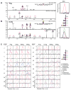
Fig. 2. Lfng, Mfng, and Rfng generate unique patterns of O-fucose glycoforms on N1 EGF1-36
Peptides derived from mouse N1 EGF1-36 produced in the absence (A) or presence (B) of Lfng were analyzed by mass spectrometry. The top panels show an MS spectrum at 4.5 min, and the bottom panels are the MS/MS spectrum of the indicated glycopeptide from EGF35, confirming the assignment. An Extracted Ion Chromatogram (EIC) of the MS data following the relative amounts of the ions corresponding to the potential glycoforms of this peptide (unmodified, mono-, di-, tri- and tetra-saccharide glycoforms, see key) is shown on the right. (C) EICs were generated as in (A) and (B) to evaluate the relative amounts of the O-fucose glycoforms (see key on right) on each O-fucosylated peptide. Spectra supporting the assignments of the ions used to generate the EICs for each site can be found in Data S1. Several of the peptides have additional modifications (see footnotes). Key: black bar, peptide; red triangle, fucose; blue square, GlcNAc; yellow circle, galactose; purple diamond, sialic acid.
Fig. 3
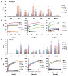
Fig. 3. Fringe-mediated elongation of O-fucose at specific EGF repeats modulates N1 activation from DLL1 or J1
(A) WT N1 or O-fucosylation site mutants (or Empty Vector, EV) were tested in cell-based N1 activation assays using DLL1 as activating ligand. Assays were performed as indicated in the absence (blue) or presence of Lfng (red), Mfng (green) or Rfng (purple). Analysis of additional mutants is shown in Fig. S3D. Bar graph shows mean +/− SD; Statistical significance of the enhancement of activation relative to –Fng for each mutant was determined using one-way ANOVA. Three independent experiments n = 9 were analyzed. ***, p < 0.0001; **, p < 0.001; *, P < 0.01. (B) Cell-based N1-DLL1 binding assays at a fixed concentration of DLL1 were performed. HEK293T cells were co-transfected with WT or mutant N1 (or EV) and GFP along with increasing amounts of Lfng (left), Mfng (middle), or Rfng (right). Cells were incubated with 3.6 nM DLL1-Fc pre-incubated with PE-Anti-mouse IgG as described in Supplemental Experimental Procedures. (C). N1 activation assays were done as in panel A but J1 was used as activating ligand. Analysis of additional mutants is shown in Fig. S3E. (D). N1-J1 binding assays at a fixed concentration of J1 were performed as in panel B. Cells were incubated with 2.1 nM J1-Fc pre-incubated with PE-Anti-human IgG as described in Supplemental Experimental Procedures.
Fig. 4
Fig. 4. The Fringe-mediated Notch Code
A summary of the data from Fig. 2C. The most abundant O-fucose glycan found at each site is diagrammed. Fringe modification at EGF12 enhances binding to and activation of N1 by both ligands (green arrow), while modification at EGF8 enhances binding and activation from DLL1 (blue arrow). Fringe modifications at EGF6 and 36 inhibit N1 activation from J1 even though binding is increased due to modification at EGF12 (red arrows).

Similar articles

Cited by

References

    1. Andrawes MB, Xu X, Liu H, Ficarro SB, Marto JA, Aster JC, Blacklow SC. Intrinsic Selectivity of Notch 1 for Delta-like 4 over Delta-like 1. J Biol Chem. 2013;288:25477–25489. - PMC - PubMed
    1. Benedito R, Roca C, Sorensen I, Adams S, Gossler A, Fruttiger M, Adams RH. The notch ligands Dll4 and Jagged1 have opposing effects on angiogenesis. Cell. 2009;137:1124–1135. - PubMed
    1. Bruckner K, Perez L, Clausen H, Cohen S. Glycosyltransferase activity of Fringe modulates Notch-Delta interactions. Nature. 2000;406:411–415. - PubMed
    1. Chen J, Moloney DJ, Stanley P. Fringe modulation of Jagged1-induced Notch signaling requires the action of ß4Galactosyltransferase-1. Proc Natl Acad Sci USA. 2001;98:13716–13721. - PMC - PubMed
    1. D'Amato G, Luxan G, Del Monte-Nieto G, Martinez-Poveda B, Torroja C, Walter W, Bochter MS, Benedito R, Cole S, Martinez F, et al. Sequential Notch activation regulates ventricular chamber development. Nat Cell Biol. 2016;18:7–20. - PMC - PubMed

MeSH terms