. 2017 Jan 19;65(2):336-346.
doi: 10.1016/j.molcel.2016.12.007. Epub 2017 Jan 12.

Coupling of Homologous Recombination and the Checkpoint by ATR

Affiliations

Coupling of Homologous Recombination and the Checkpoint by ATR

Rémi Buisson et al. Mol Cell. .

Abstract

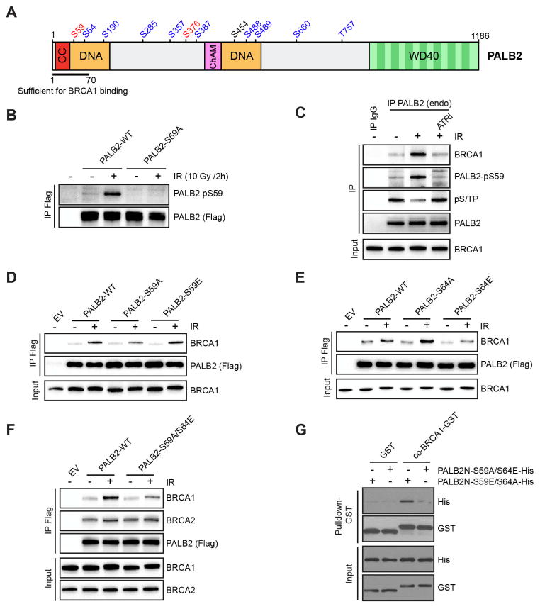
ATR is a key regulator of cell-cycle checkpoints and homologous recombination (HR). Paradoxically, ATR inhibits CDKs during checkpoint responses, but CDK activity is required for efficient HR. Here, we show that ATR promotes HR after CDK-driven DNA end resection. ATR stimulates the BRCA1-PALB2 interaction after DNA damage and promotes PALB2 localization to DNA damage sites. ATR enhances BRCA1-PALB2 binding at least in part by inhibiting CDKs. The optimal interaction of BRCA1 and PALB2 requires phosphorylation of PALB2 at S59, an ATR site, and hypo-phosphorylation of S64, a CDK site. The PALB2-S59A/S64E mutant is defective for localization to DNA damage sites and HR, whereas the PALB2-S59E/S64A mutant partially bypasses ATR for its localization. Thus, HR is a biphasic process requiring both high-CDK and low-CDK periods. As exemplified by the regulation of PALB2 by ATR, ATR promotes HR by orchestrating a "CDK-to-ATR switch" post-resection, directly coupling the checkpoint to HR.

Keywords: ATR; BRCA1; CDK; PALB2; checkpoint; homologous recombination.


Figures

Fig. 1
Fig. 1. ATR promotes PALB2 localization to DSBs after resection
A. BRCA1 and RAD51 localization in U2OS cells 2 h after IR (4 Gy). Cells were pre-treated with DMSO or ATRi for 30 min before IR. B. Quantification of cells displaying >5 RAD51 foci in A. Error bar: S.D. (n=3). C. RPA and RAD51 localization in HeLa cells 4 h after IR (10 Gy). Cells were pre-treated with DMSO of ATRi 30 min before IR. D. BRCA1 and PALB2 localization in U2OS cells 2 h after IR (4 Gy). Cells were pre-treated with DMSO or ATRi 30 min before IR. E. Quantification of cells displaying >10 BRCA1 foci in D. Error bar: S.D. (n=3). F. The fractions of BRCA1 foci-positive cells displaying >10 PALB2 foci were quantified after DMSO or ATRi treatment. Error bar: S.D. (n=3). G. Localization of YFP-PALB2WT to laser-induced DNA damage sites was analyzed by time-lapse after indicated treatment. The fluorescence intensity of YFP-PALB2WT at DNA damage sites was quantified over time. For each cell analyzed, the signal in an unirradiated nuclear area in the same cell was determined as background. Error bar: S.E. (n > 50).
Fig. 2
Fig. 2. ATR promotes the BRCA1-PALB2 interaction through CDK inhibition and an additional mechanism
A. Immunoprecipitation of PALB2 from HeLa cells after IR (10 Gy) at the indicated time points. Co-immunoprecipitated BRCA1 was analyzed by Western blot. B. HeLa cells were treated with ATRi or Chk1i prior to IR (10 Gy). PALB2 was immunoprecipitated 2 h after IR, and levels of co-immunoprecipitated BRCA1 and BRCA2 were analyzed by Western blot. C. HeLa cells were treated with ATR inhibitor prior to IR (4 Gy). PALB2-ΔETGE was immunoprecipitated 2 h after IR, and levels of co-immunoprecipitated BRCA1 were analyzed by Western blot. D. HeLa cells were irradiated (10 Gy) and levels of the indicated proteins were analyzed by Western blot at the shown time points. E. HeLa cells were treated with ATRi prior to IR (4 Gy). Endogenous PALB2 was immunoprecipitated and levels of pS/TP were analyzed by Western blot. F. HeLa cells were treated with Wee1 inhibitor prior to IR (10 Gy). PALB2 was immunoprecipitated 2 h after IR, and levels of co-immunoprecipitated BRCA1 were analyzed by Western blot. G-I. U2OS cells were treated with DMSO or ATRi 1 h before IR, and then with DMSO, Roscovitine, or PHA-793887 1 h after IR. Fractions of BRCA1 foci-positive cells displaying >10 PALB2 foci (H) or >5 RAD51 foci (I) were quantified. Error bar: S.D. (n=2 in H, n=3 in I)
Fig. 3
Fig. 3. PALB2 phosphorylation at S59 and S64 affects BRCA1 binding
A. A schematic representation of PALB2 and the 12 phosphorylation sites identified by mass spectrometry. S/TQ motifs are shown in red and S/TP motifs are shown in blue. CC : Coil –coiled domain; DNA: DNA binding domain; ChAM: Chromatin-Association Motif; WD40: WD40-repeat-containing domain. B. Flag-tagged PALB2WT or PALB2S59A were transiently expressed in HeLa cells. Flag immunoprecipitation was performed 2 h after IR (10 Gy), and levels of pS59 were analyzed by Western blot. C. HeLa cells were treated with ATRi prior to IR (4 Gy). At 4 h after irradiation, endogenous PALB2 was immunoprecipitated, and levels of pS59, pS/TP, and co-immunoprecipitated BRCA1 were analyzed by Western blot. D-F. Indicated Flag-tagged PALB2 variants were transiently expressed in HeLa cells. Flag immunoprecipitation was performed 2 h after IR, and levels of co-immunoprecipitated BRCA1 or BRCA2 were analyzed by Western blot. G. Purified GST-tagged BRCA1 coiled-coil domain (BRCA1-cc) was incubated with purified His-tagged PALB2N fragments (PALB2N) containing the indicated mutations. The direct interaction between BRCA1-cc and PALB2N was analyzed by GST pulldown.
Fig. 4
Fig. 4. The S59/S64 phosphorylation switch on PALB2 regulates its localization and HR function
A. Localization of YFP-PALB2 variants to laser-induced DNA damage sites was analyzed by time-lapse. The fluorescence intensity of YFP-PALB2 at DNA damage sites was quantified as in Fig. 1G. Error bar: S.E. (n > 50). B. Indicated Flag-tagged PALB2 variants were transiently expressed in HeLa cells. Cells were treated DMSO or ATRi, irradiated with IR, and Flag immunoprecipitation was performed 2 h after IR. Levels of co-immunoprecipitated BRCA1 were analyzed by Western blot. C. Representative images of GFP-PALB2WT or GFP-PALB2S59E/S64A foci in DMSO or ATRi-treated cells 2 h after IR. D. The intensity of PALB2 staining in BRCA1 foci was determined in the indicated cell populations. Cells expressing GFP-tagged PALB2WT, PALB2S59E/S64A, or PALB2S59A/S64E were treated with DMSO or ATRi as shown. ***, P< 0.001 and ****, P< 0.0001. E. Localization of YFP-PALB2 variants to laser-induced DNA damage sites in the presence of ATRi (1 μM VE-821). Error bar: S.E. (n > 50). F-G. Flag-tagged PALB2 variants were stably integrated in U2OS cells under the control of a Tet-on promoter. Cells were treated with PALB2 siRNA to deplete endogenous PALB2, and with Doxycycline to induce the expression of the PALB2 variants. Gene-targeting efficiency were quantified (mClover positive cells) after the indicated treatments. *, P<0.05; **, P< 0.01 and ****, P< 0.0001.

References

    1. Adamson B, Smogorzewska A, Sigoillot FD, King RW, Elledge SJ. A genome-wide homologous recombination screen identifies the RNA-binding protein RBMX as a component of the DNA-damage response. Nat Cell Biol. 2012;14:318–328. - PMC - PubMed
    1. Ahlskog JK, Larsen BD, Achanta K, Sorensen CS. ATM/ATR-mediated phosphorylation of PALB2 promotes RAD51 function. EMBO Rep. 2016;17:671–681. - PMC - PubMed
    1. Buisson R, Dion-Cote AM, Coulombe Y, Launay H, Cai H, Stasiak AZ, Stasiak A, Xia B, Masson JY. Cooperation of breast cancer proteins PALB2 and piccolo BRCA2 in stimulating homologous recombination. Nat Struct Mol Biol. 2010;17:1247–1254. - PMC - PubMed
    1. Buisson R, Masson JY. PALB2 self-interaction controls homologous recombination. Nucleic Acids Res. 2012;40:10312–10323. - PMC - PubMed
    1. Busino L, Donzelli M, Chiesa M, Guardavaccaro D, Ganoth D, Dorrello NV, Hershko A, Pagano M, Draetta GF. Degradation of Cdc25A by beta-TrCP during S phase and in response to DNA damage. Nature. 2003;426:87–91. - PubMed

MeSH terms