. 2017 Jan 16:8:14017.
doi: 10.1038/ncomms14017.

A mast cell-ILC2-Th9 pathway promotes lung inflammation in cystic fibrosis

Affiliations

A mast cell-ILC2-Th9 pathway promotes lung inflammation in cystic fibrosis

Silvia Moretti et al. Nat Commun. .

Abstract

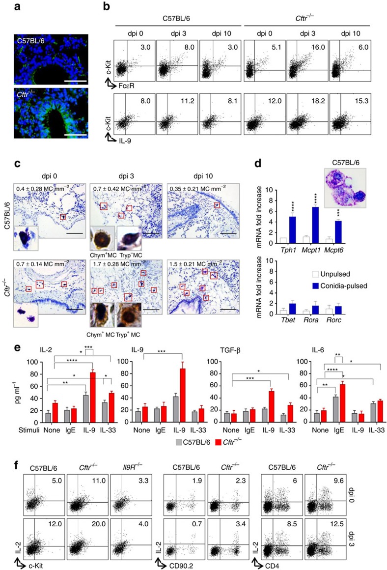
T helper 9 (Th9) cells contribute to lung inflammation and allergy as sources of interleukin-9 (IL-9). However, the mechanisms by which IL-9/Th9 mediate immunopathology in the lung are unknown. Here we report an IL-9-driven positive feedback loop that reinforces allergic inflammation. We show that IL-9 increases IL-2 production by mast cells, which leads to expansion of CD25+ type 2 innate lymphoid cells (ILC2) and subsequent activation of Th9 cells. Blocking IL-9 or inhibiting CD117 (c-Kit) signalling counteracts the pathogenic effect of the described IL-9-mast cell-IL-2 signalling axis. Overproduction of IL-9 is observed in expectorates from cystic fibrosis (CF) patients, and a sex-specific variant of IL-9 is predictive of allergic reactions in female patients. Our results suggest that blocking IL-9 may be a therapeutic strategy to ameliorate inflammation associated with microbial colonization in the lung, and offers a plausible explanation for gender differences in clinical outcomes of patients with CF.


Figures

Figure 1
Figure 1. IL-9 production and ILC2-Th9 cells activation in Aspergillus fumigatus infection.
(a) Time course of IL-9 production at various days post infection (dpi) in mice (six per group) infected intranasally with live A. fumigatus conidia. (b) Detection of CD90.2+CD25+, CD90.2+ST2+ and CD90.2+IL-9+ lung type 2 ILCs by flow cytometry (numbers refer to percentages of positive cells) and immunofluorescence staining. (c) Absolute number of lung ILC2; (d) ILC2–specific transcript on lineage negative lung cells; (e,f) ILC2 effector and activating cytokines; (g) Il9 and Th9-cell specific transcripts on lung CD4+ T cells and (h) immunofluorescence staining of lung CD4+ IL-9+ T cells. Photographs were taken with a high-resolution microscope (Olympus DP71) equipped with a × 40 objective; scale bar, 100 μm. Mean values±s.d. cytokines were determined on lung homogenates by ELISA, Il9 and transcripts assessed by PCR with reverse transcription. 0, uninfected mice. *P<0.05, **P<0.01, ***P<0.001, ****P<0.0001, knockout versus C57BL/6 mice (data represent pooled results or representative images from three experiments, Two-way ANOVA, Bonferroni post test). Gata3, GATA binding protein 3; Irf4, interferon regulatory factor 4; Pu.1, purine-rich box 1; Rora, RAR-related orphan receptor alpha.
Figure 2
Figure 2. IL-9R signaling contributes to inflammation and allergy.
C57BL/6 and Il9R−/− mice (six per group) were intranasally infected with live Aspergillus fumigatus conidia or subjected to ABPA and assessed for (a) lung fungal growth (log10 cfu, mean±s.d.); (b) lung histology (periodic acid−Schiff staining); (c) expression of CD90.2+CD25+, CD90.2+ST2+ lung ILC2 by immunofluorescence; (d,e) Th-cell specific transcripts and cytokine production. (f) Lung histology (periodic acid–Schiff and, in the inset, Masson's trichrome staining) and (g) TGF-β production in C57BL/6 or Cftr−/− mice infected as above and treated with IL-9 neutralizing antibody for a week. Days post infection (dpi). (h) Th-cell specific transcripts and IL-9 production of lung CD4+ T cells from naive mice co-cultured with lung lineage negative (Lin) cells in the presence of A. fumigatus conidia, IL-2 or IL-33. Photographs were taken with a high-resolution microscope (Olympus DP71) equipped with a × 20 objective; scale bars, 200 μm and a × 40 objective (insets of f, scale bars, 100 μm). Results are mean values±s.d., ELISA was done on lung homogenates and culture supernatants for cytokines and PCR with reverse transcription on CD4+ lung cells. *P<0.05, **P<0.01, ***P<0.001, Il9R−/−, Cftr−/− versus C57BL/6 mice; IL-9-treated versus control isotype-treated mice; stimulated versus unstimulated (none) cells and Il9R−/− versus C57BL/6 or Cftr−/− CD4+ T cells. Naive, uninfected mice. Data represent pooled results or representative images from three experiments, Two-tailed Student's t-test (a) or Two-way ANOVA (d,e) Bonferroni post test. Gata3, GATA binding protein 3; Irf4, interferon regulatory factor 4; Pu.1, purine-rich box 1.
Figure 3
Figure 3. IL-9 activates mast cells to produce IL-2.
C57BL/6 or Cftr−/− mice (six per group) infected intranasally with live A. fumigatus conidia were evaluated at different days after infection (dpi) for (a) deposition of DNA on lung epithelial cells by TUNEL, resulting in bright DNA staining; (b) detection of c-Kit+FcɛR+ and c-Kit+IL-9+ lung mast cells (MC) by flow cytometry (numbers refer to percentages of positive cells); (c) toluidine blue, relative MC number mm−2 and, in the inset, immunohistochemical staining for chymase- and tryptase-positive MC in lung section. Photographs were taken with a high-resolution microscope (Olympus DP71) equipped with a × 40 objective and (in the inset) a × 100 objective and with EVOS FL Color Imaging System with a × 60 objective (immunohistochemical staining). (d) Toluidine blue stain and transcription factors expression (PCR with reverse transcription) of c-Kit+ cells magnetically isolated from lung of uninfected C57BL/6 mice and pulsed with live A. fumigatus conidia. (e) Cytokine production (mean values±s.d., ELISA on culture supernatants) by purified lung c-Kit+ cells, pulsed with A. fumigatus and stimulated with IgE, IL-9 and IL-33; (f) detection of c-Kit+IL-2+, CD90.2+IL-2+ and CD4+IL-2+ lung cells by flow cytometry (numbers refer to percentages of positive cells). *P<0.05, **P<0.01, ***P<0.001, ****P<0.0001, conidia-pulsed versus unpulsed c-Kit+ cells, stimulated versus unstimulated c-Kit+ cells and Cftr−/− versus C57BL/6 c-Kit+ cells (data represent pooled results or representative images from three experiments, Two-way ANOVA, Bonferroni post test). Mcpt1, mast cell protease 1; Mcpt6, tryptase beta 2; Rorc, retinoic acid receptor–related orphan receptor C; Rora, RAR-related orphan receptor alpha; Tbet, T box expressed in T cells; Tph1, tryptophan hydroxylase 1.
Figure 4
Figure 4. IL-9 activates mast cells to produce IL-2.
MC-deficient C57BL6-KitW/W-v mice (six per group) were infected intranasally with live A. fumigatus conidia, engrafted intravenously with wild-type bone marrow-cultured mast cells (BMMC) or treated intraperitoneally with IL-2 for a week and assessed for (a) c-Kit+FcɛR+ lung mast cells (MC) and CD90.2+CD25+ lung ILC2 by flow cytometry (numbers refer to percentages of positive cells) with relative cell number and (b) IgE and cytokine production. (c) Lung histology (periodic acid–Schiff and Masson's trichrome staining, in the insets); (d) cytokine production and (e) Th9-cell specific transcripts expression in Cftr−/− mice infected as above and treated with imatinib intraperitoneally for a week. Photographs were taken with a high-resolution microscope (Olympus DP71) equipped with a × 20 objective, scale bars, 200 μm and a × 40 objective (insets of c, scale bars, 100 μm). Results are mean values±s.d., ELISA on lung homogenates for cytokines and PCR with reverse transcription on lung CD4+T cells. *P<0.05, **P<0.01, ***P<0.001, ****P<0.0001, MC-deficient C57BL6-KitW/W-v versus C57BL/6 mice or imatinib-treated versus untreated (none) mice (data represent pooled results or representative images from three experiments, Two-way ANOVA, Bonferroni post test). Irf4=interferon regulatory factor 4; Pu.1=purine-rich box 1.
Figure 5
Figure 5. Epithelial and myeloid CFTR deficiency contribute to the inflammatory phenotype.
C57BL/6, Cftr−/− and chimeric C57BL/6 and Cftr−/− mice (10 per group) received 10 × 106 viable bone marrow cells 4 weeks before the intranasal infection with A. fumigatus. Chimeric mice were evaluated 7 days after the infection for (a) lung histology (periodic acid–Schiff and, in the insets, Masson's trichrome and TUNEL staining); (b) cytokines and IgE levels (mean values±s.d., ELISA on lung homogenates); (c) detection of c-Kit+FcɛR+ mast cells, CD90.2+CD25+ and CD90.2+ST2+ type 2 ILCs by flow cytometry (numbers refer to percentages of positive cells in the lung). Photographs were taken with a high-resolution microscope (Olympus DP71) equipped with a × 20 objective; scale bars, 200 μm and a × 40 objective (insets of panel a, scale bars, 100 μm). *P<0.05, **P<0.01, ***P<0.001, Cftr−/− versus C57BL/6, chimeric C57BL/6 versus C57BL/6, chimeric Cftr−/− versus Cftr−/− mice, Two-way ANOVA, Bonferroni post test.
Figure 6
Figure 6. The IL9 rs2069885 polymorphism correlates with high IgE levels in CF females.
(a,b) IL9 rs2069885 sex interaction on total or Aspergillus-specific IgE levels and (c) IL9/IL9R expression ratio measured on CF patients. (d) Determination IL-9 (mean±s.d., ELISA) in expectorates from CF patients carrying diverse genotypes at rs2069885.
Figure 7
Figure 7. Proposed model for the role of IL-9 in promoting a mast cells/ILC2/Th9 fibrotic pathway in CF.
IL-9, produced by IL-33-expanded ILC2, activates MC for IL-2 production leading to the expansion of CD25+ILC2 that promote Th9 cell activation. The resulting increased production of IL-9 further amplifies the inflammatory loop by promoting ILC2 survival and type 2 cytokines production and by activating MC for the production of fibrotic TGF-β. IL-9 ablation or MC inhibition (imatinib) are potential drugable pathways through which inflammation and allergy could be restrained in CF. EC, epithelial cells. αIL-9, IL-9 neutralizing antibody.

References

    1. Spits H. et al. Innate lymphoid cells–a proposal for uniform nomenclature. Nat. Rev. Immunol. 13, 145–149 (2013). - PubMed
    1. Roediger B. & Weninger W. Group 2 innate lymphoid cells in the regulation of immune responses. Adv. Immunol. 125, 111–154 (2015). - PubMed
    1. Lund S., Walford H. H. & Doherty T. A. Type 2 innate lymphoid cells in allergic disease. Curr. Immunol. Rev. 9, 214–221 (2013). - PMC - PubMed
    1. Drake L. Y. & Kita H. Group 2 innate lymphoid cells in the lung. Adv. Immunol. 124, 1–16 (2014). - PMC - PubMed
    1. Halim T. Y., Krauss R. H., Sun A. C. & Takei F. Lung natural helper cells are a critical source of Th2 cell-type cytokines in protease allergen-induced airway inflammation. Immunity 36, 451–463 (2012). - PubMed

Publication types

MeSH terms

Substances