. 2018 Feb;15(2):171-181.
doi: 10.1038/cmi.2016.60. Epub 2017 Jan 16.

uPAR promotes tumor-like biologic behaviors of fibroblast-like synoviocytes through PI3K/Akt signaling pathway in patients with rheumatoid arthritis

Affiliations

uPAR promotes tumor-like biologic behaviors of fibroblast-like synoviocytes through PI3K/Akt signaling pathway in patients with rheumatoid arthritis

Yan Liu et al. Cell Mol Immunol. 2018 Feb.

Abstract

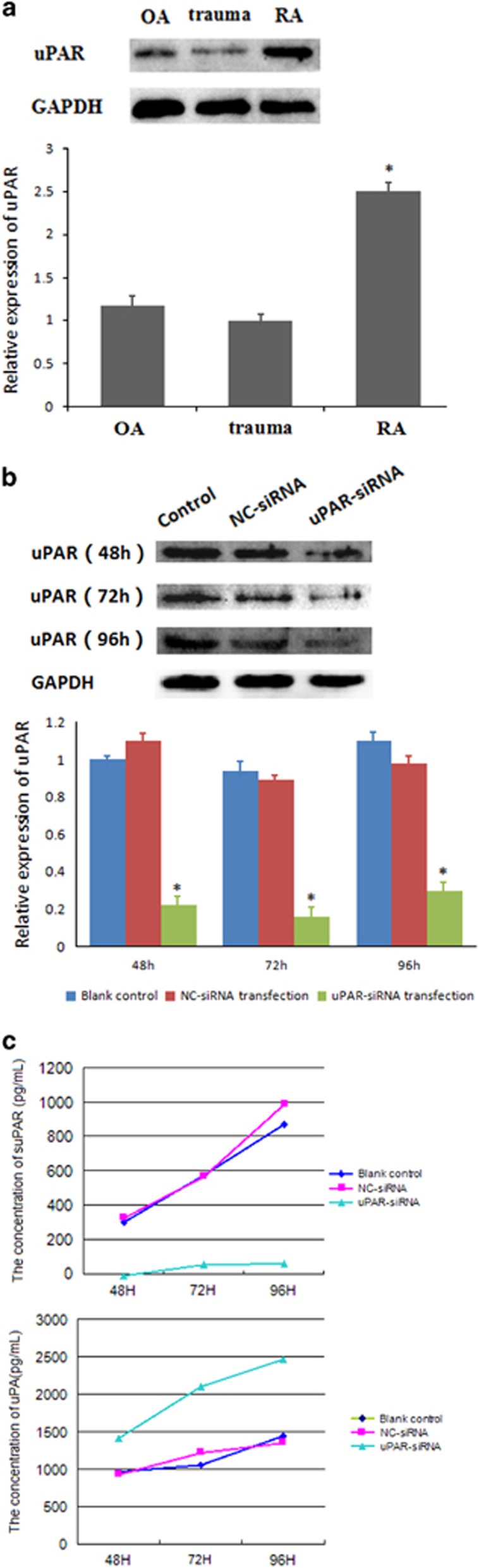
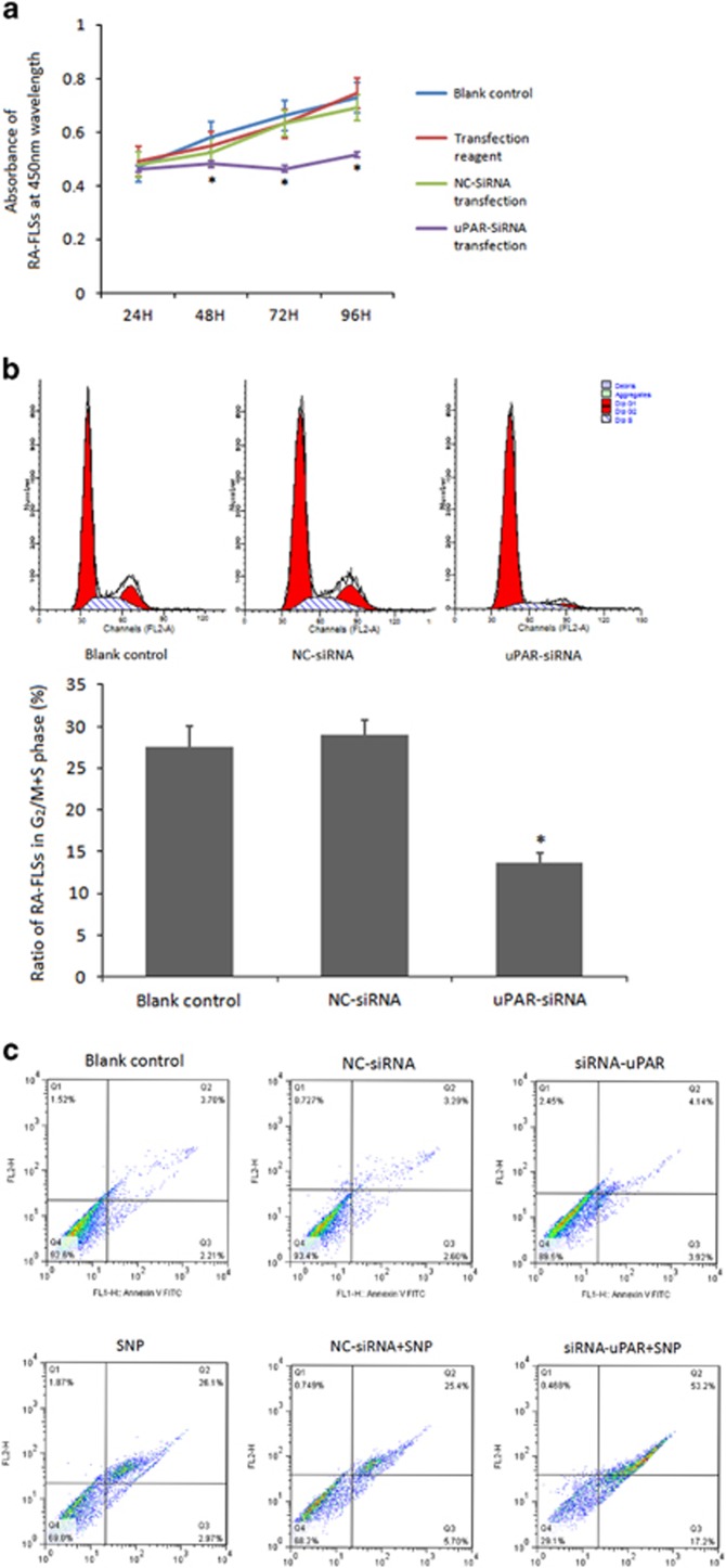
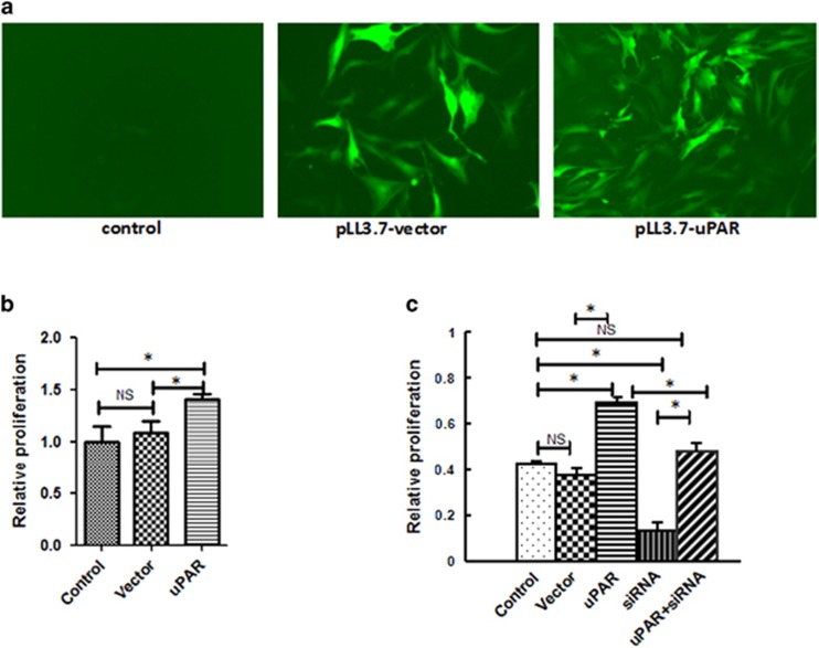
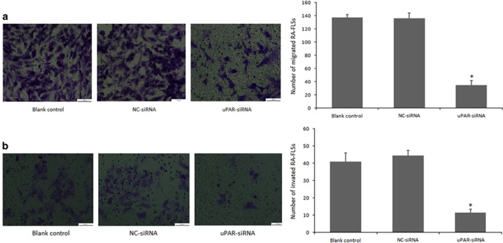
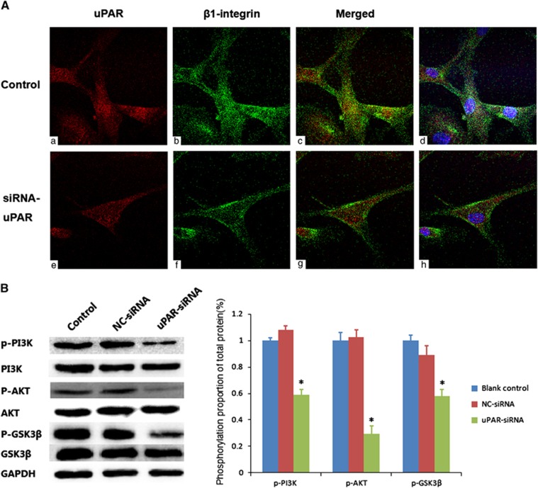
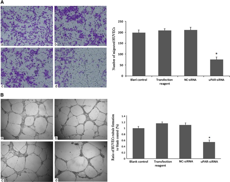
Urokinase-type plasminogen activator receptor (uPAR), is a multifunctional receptor on cell surface, widely present in endothelial cells, fibroblasts, and a variety of malignant cells. Current studies have suggested that uPAR overexpressed on synovial tissues or in synovial fluid or plasma in patients with rheumatoid arthritis (RA). However, there are limited researches regarding the role of uPAR on fibroblast-like synoviocytes of rheumatoid arthritis (RA-FLSs) and its underlying mechanisms. Here, our studies show that the expression of uPAR protein was significantly higher in fibroblast-like synoviocytes (FLSs) from RA than those from osteoarthritis or traumatic injury patients. uPAR gene silencing significantly inhibited RA-FLSs cell proliferation, restrained cell transformation from the G0/G1 phase to S phase, aggravated cell apoptosis, interfered with RA-FLSs cell migration and invasion, and reduced activation of the PI3K/Akt signaling pathway, which may be associated with β1-integrin. Cell supernatants from uPAR gene-silenced RA-FLSs markedly inhibited the migration and tubule formation ability of the HUVECs (a human endothelial cell line). Therefore, we demonstrate that uPAR changes the biological characteristics of RA-FLSs, and affects neoangiogenesis of synovial tissues in patients with RA. All of these may be associated with the β1-integrin/PI3K/Akt signaling pathway. These results imply that targeting uPAR and its downstream signal pathway may provide therapeutic effects in RA.


Conflict of interest statement

The authors declare no conflict of interest.

Figures

Figure 1
Figure 1
(a) RA-FLSs expressed higher levels of uPAR than OA and trauma groups. Western blot assays were used to detect uPAR expression in RA, OA, and traumatic FLSs. Densitometry analysis of protein expression (means±s.e.m.) were representative of three independent experiments. (b) uPAR-siRNA efficiently interferes with uPAR expression in RA-FLSs. Cells were transfected with uPAR-siRNA (40 nM) or a negative control (NC, 40 nM) for 48, 72, 96 h and western blot analysis was used to detect the effect of uPAR-siRNA on uPAR expression. Upper panel, representative images are shown; lower panel, quantification (means±s.e.m.) of three independent experiments normalized to 1 in the untreated sample. (c) uPAR knockdown affects suPAR/uPA secretion. Secretion curves of suPAR/uPA in the supernatants of transfected RA-FLSs for 48 , 72 , 96 h was made by ELISA assays of three independent experiments. *P<0.05, compared with the controls and n=4. FLSs, fibroblast-like synoviocytes; OA, osteoarthritis; uPAR, Urokinase-type plasminogen activator receptor.
Figure 2
Figure 2
(a) Knocking-down uPAR in RA-FLSs decreases cell proliferation. RA-FLSs were transfected with uPAR-siRNA (40 nM) or a negative control (NC, 40 nM) for 24 h, 48 h, 72 h, 96 h, respectively, and cell proliferation was assessed by the CCK-8 assay. (b) uPAR knockdown in RA-FLSs influences cell cycle. Flow cytometry analysis was performed to analyze the effects of uPAR on cell-cycle distribution. The upper panel was a representative histogram, the lower panel is the cumulative data of the effects. (c) uPAR knockdown in RA-FLSs aggravates cell apoptosis. After treatment of apoptosis inducer SNP and transfection, flow cytometry assay was also used to detect the RA-FLSs cell apoptotic rates. Annexin-V-FITC(+)PI(+) and Annexin-V-FITC(+)PI(−) population represented apoptotic cells. *P<0.05, compared with the controls and n=4. RA-FLSs, fibroblast-like synoviocytes of rheumatoid arthritis; uPAR, Urokinase-type plasminogen activator receptor.
Figure 3
Figure 3
Lentivirus-mediated uPAR overexpression promotes RA-FLS cells proliferation. Structures of pLentilox 3.7 contains GFP reporter gene. (a) Transfection efficiency of lentivirus-mediated overexpression in FLS cells was evaluated by GFP signal under fluorescence microscope (magnification × 100). (b) CCK-8 analysis showed that lentivirus-mediated uPAR overexpression significantly promotes RA-FLS cells proliferation. (c) Rescued experiment revealed that lentivirus-mediated uPAR overexpression reverses inhibition effects by uPAR-siRNA (c). *P<0.05, compared with the controls and n=4. RA-FLSs, fibroblast-like synoviocytes of rheumatoid arthritis; uPAR, Urokinase-type plasminogen activator receptor.
Figure 4
Figure 4
Cell migration and invasion ability is impaired after uPAR knockdown in RA-FLSs. In both (a) and (b), RA-FLSs were transfected with uPAR-siRNA (40 nM) or a negative control (NC, 40 nM) for 72 h. (a) The RA-FLSs migration was measured in a transwell chamber assay. (b) The RA-FLSs invasive ability was assessed by a Matrigel invasion assay. In both (a) and (b), left panels show representative images; right panels present quantification (means±s.e.m.) of three independent experiments. *P<0.05, compared with blank control and the NC-siRNA control and n=4. RA-FLSs, fibroblast-like synoviocytes of rheumatoid arthritis; uPAR, Urokinase-type plasminogen activator receptor.
Figure 5
Figure 5
(A) uPAR co-locates with β1-integrin and interferes its internalization. To facilitate visualization, images were converted to pseudo color using ImageJ software: uPAR (red, a, e), β1-integrin (green, b, f), and sites of colocalization (yellow, c, g). The nuclear staining was added in the merged images for easy location and shown on the right (blue, d, h). The above experiments were performed three times with similar results. (B) uPAR silencing reduces PI3K/Akt downstream signaling activation in RA-FLSs. Western blot showed the levels of phosphorylated PI3K, Akt and GSK3β in RA-FLSs after uPAR-siRNA (40 nM) or negative control (NC, 40 nM) following transfection for 72 h. Left panels show representative protein strips; right panels present densitometry quantification of protein expression (means±s.e.m.) of three independent experiments. *P<0.05, compared with the controls and n=4. RA-FLSs, fibroblast-like synoviocytes of rheumatoid arthritis; uPAR, Urokinase-type plasminogen activator receptor.
Figure 6
Figure 6
Interference of uPAR in RA-FLSs suppresses endotheliocyte angiopoiesis by reducing cell migration and tubule formation. (A) The HUVECs cell migration ability in different conditioned media were evaluated by a transwell chamber assay. (a) Blank control group, serum-free DMEM; (b) Transfection reagent group, conditioned medium of RA-FLSs transfected only with transfection reagent; (c) NC-siRNA group, conditioned medium of RA-FLSs transfected with a negative control (NC, 40 nM); (d) uPAR-siRNA group, conditioned medium of RA-FLSs transfected with uPAR-siRNA (40 nM). (B) The HUVECs cells with different conditioned media were added into Matrigel coated 96-well for 24 h to form tubule. Conditioned media treatment just reference as A. In both (A) and (B), left panels show representative images; right panels present quantification of tubule length ratio to controls (means±s.e.m.) of three independent experiments. *P<0.05, compared with blank control and n=4. HUVECs, human endothelial cell lines; RA-FLSs, fibroblast-like synoviocytes of rheumatoid arthritis; uPAR, Urokinase-type plasminogen activator receptor.

References

    1. Henry J, Roulot E, Gaujoux-Viala C. The rheumatoid hand. Presse Med 2013; 42: 1607–1615. - PubMed
    1. Bartok B, Firestein GS. Fibroblast-like synoviocytes: key effector cells in rheumatoid arthritis. Immunol Rev 2010; 233: 233–255. - PMC - PubMed
    1. Huber LC, Distler O, Tarner I, Gay RE, Gay S, Pap T. Synovial fibroblasts: key players in rheumatoid arthritis. Rheumatology 2006; 45: 669–675. - PubMed
    1. Rao JS, Gujrati M, Chetty C. Tumor-associated soluble uPAR-directed endothelial cell motility and tumor angiogenesis. Oncogenesis 2013; 2: e53. - PMC - PubMed
    1. Shetty S, Kumar A, Johnson AR, Pueblitz S, Holiday D, Raghu G et al. Differential expression of the urokinase receptor in fibroblasts from normal and fibrotic human lungs. Am J Respir Cell Mol Biol 1996; 15: 78–87. - PubMed

Publication types

MeSH terms

Substances