

Multimodality Imaging for Left Ventricular Hypertrophy Severity Grading: A Methodological Review
- PMID: 28090249
- PMCID: PMC5234336
- DOI: 10.4250/jcu.2016.24.4.257
Multimodality Imaging for Left Ventricular Hypertrophy Severity Grading: A Methodological Review
Abstract
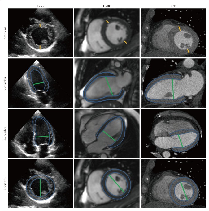
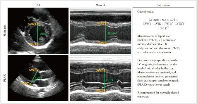
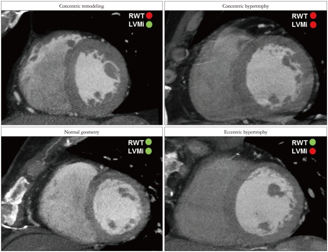
Left ventricular hypertrophy (LVH), defined by an increase in left ventricular mass (LVM), is a common cardiac finding generally caused by an increase in pressure or volume load. Assessing severity of LVH is of great clinical value in terms of prognosis and treatment choices, as LVH severity grades correlate with the risk for presenting cardiovascular events. The three main cardiac parameters for the assessment of LVH are wall thickness, LVM, and LV geometry. Echocardiography, with large availability and low cost, is the technique of choice for their assessment. Consequently, reference values for LVH severity in clinical guidelines are based on this technique. However, cardiac magnetic resonance (CMR) and computed tomography (CT) are increasingly used in clinical practice, providing excellent image quality. Nevertheless, there is no extensive data to support reference values based on these techniques, while comparative studies between the three techniques show different results in wall thickness and LVM measurements. In this paper, we provide an overview of the different methodologies used to assess LVH severity with echocardiography, CMR and CT. We argue that establishing reference values per imaging modality, and possibly indexed to body surface area and classified per gender, ethnicity and age-group, might be essential for the correct classification of LVH severity.
Keywords: Echocardiography; Hypertrophy, left ventricular; Magnetic resonance imaging; Multidetector computed tomography.
Figures
References
-
- Cuspidi C, Sala C, Negri F, Mancia G, Morganti A Italian Society of Hypertension. Prevalence of left-ventricular hypertrophy in hypertension: an updated review of echocardiographic studies. J Hum Hypertens. 2012;26:343–349. - PubMed
-
- Ruilope LM, Schmieder RE. Left ventricular hypertrophy and clinical outcomes in hypertensive patients. Am J Hypertens. 2008;21:500–508. - PubMed
-
- Cuspidi C, Rescaldani M, Sala C, Grassi G. Left-ventricular hypertrophy and obesity: a systematic review and meta-analysis of echocardiographic studies. J Hypertens. 2014;32:16–25. - PubMed
-
- de Simone G, Izzo R, De Luca N, Gerdts E. Left ventricular geometry in obesity: is it what we expect? Nutr Metab Cardiovasc Dis. 2013;23:905–912. - PubMed
-
- Chobanian AV, Bakris GL, Black HR, Cushman WC, Green LA, Izzo JL, Jr, Jones DW, Materson BJ, Oparil S, Wright JT, Jr, Roccella EJ National Heart, Lung, and Blood Institute Joint National Committee on Prevention, Detection, Evaluation, and Treatment of High Blood Pressure; National High Blood Pressure Education Program Coordinating Committee. The Seventh Report of the Joint National Committee on Prevention, Detection, Evaluation, and Treatment of High Blood Pressure: the JNC 7 report. JAMA. 2003;289:2560–2572. - PubMed
Publication types
LinkOut - more resources
Full Text Sources
Other Literature Sources
