

Large Animal Models: The Key to Translational Discovery in Digestive Disease Research
- PMID: 28090566
- PMCID: PMC5235339
- DOI: 10.1016/j.jcmgh.2016.09.003
Large Animal Models: The Key to Translational Discovery in Digestive Disease Research
Abstract
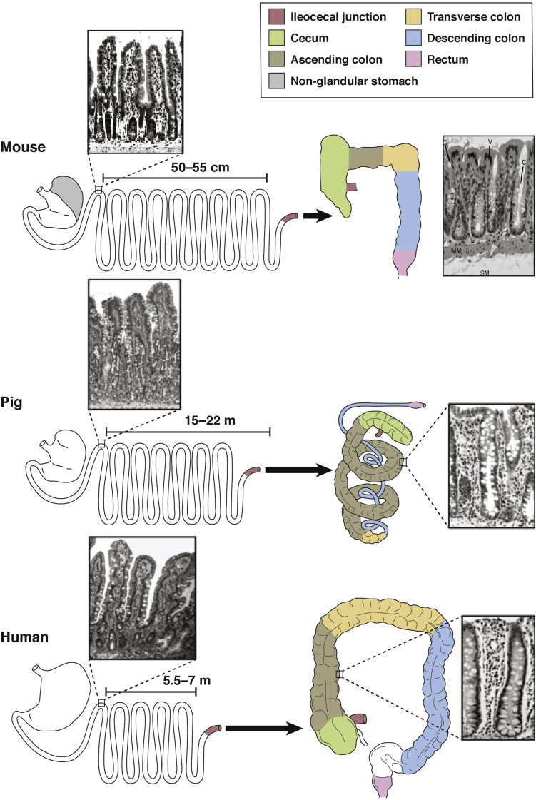
Gastrointestinal disease is a prevalent cause of morbidity and mortality and the use of animal models have been instrumental in studying mechanisms of digestive pathophysiology. As investigators attempt to translate the wealth of basic science information developed from rodent, models, large animal models provide a number of translational advantages. The pig, in particular, is arguably one of the most powerful models of human organ systems, including the gastrointestinal tract. The pig has provided important tools and insight into intestinal ischemia/reperfusion injury, intestinal mucosal repair, as well as new insights into esophageal injury and repair. Porcine model development has taken advantage of the size of the animal, allowing increased surgical and endoscopic access. In addition, cellular tools such as the intestinal porcine epithelial cell line and porcine enteroids are providing the methodology to translate basic science findings using in-depth mechanistic analyses. Further opportunities in porcine digestive disease modeling include developing additional transgenic pig strains. Collectively, porcine models hold great promise for the future of clinically relevant digestive disease research.
Keywords: Ischemia/Reperfusion Injury; Mucosal Repair; Pig; Tight Junction.
Conflict of interest statement
The authors disclose no conflicts.
Figures
References
-
- Low M.J. Mouse models in gastroenterology research. Gastroenterology. 2012;143:1410–1412. - PubMed
-
- Chung S.K., Lee A.Y., Chung S.S. Mouse models for human diseases. Hong Kong Med J. 1997;3:201–209. - PubMed
-
- Wagner S.J., Schmidt A., Effenberger M.J. Semisynthetic diet ameliorates Crohn's disease-like ileitis in TNFDeltaARE/WT mice through antigen-independent mechanisms of gluten. Inflamm Bowel Dis. 2013;19:1285–1294. - PubMed
Grants and funding
LinkOut - more resources
Full Text Sources
Other Literature Sources
Research Materials