. 2017 Jan 16:7:40213.
doi: 10.1038/srep40213.

A disrupted transsulphuration pathway results in accumulation of redox metabolites and induction of gametocytogenesis in malaria

Affiliations

A disrupted transsulphuration pathway results in accumulation of redox metabolites and induction of gametocytogenesis in malaria

Divya Beri et al. Sci Rep. .

Abstract

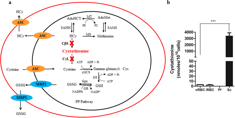
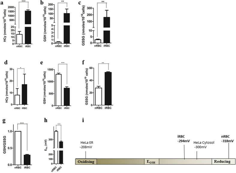
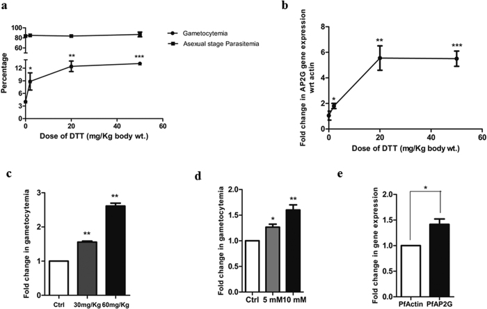
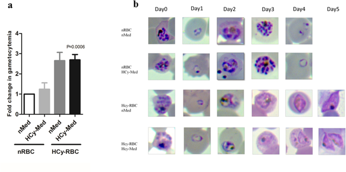
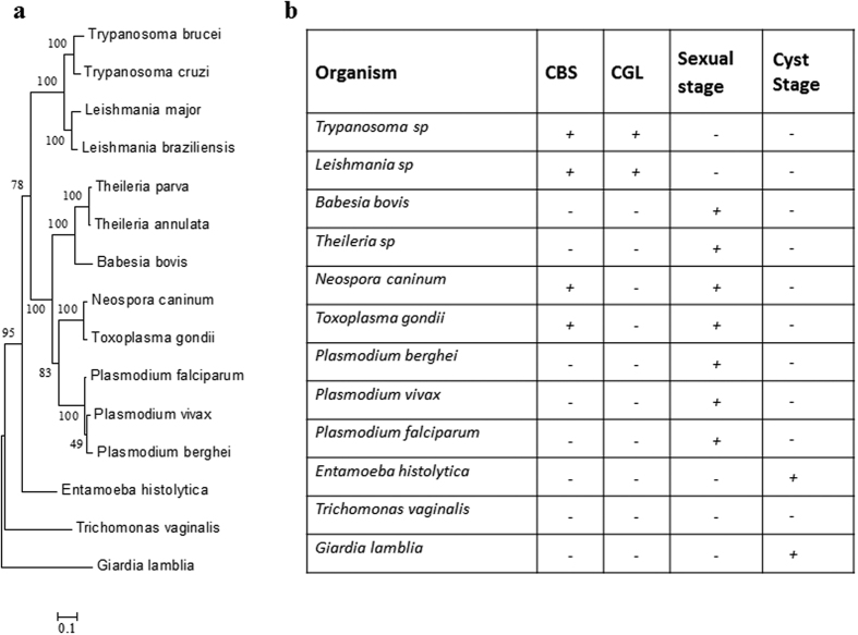
Intra-erythrocytic growth of malaria parasite is known to induce redox stress. In addition to haem degradation which generates reactive oxygen species (ROS), the parasite is also thought to efflux redox active homocysteine. To understand the basis underlying accumulation of homocysteine, we have examined the transsulphuration (TS) pathway in the parasite, which is known to convert homocysteine to cysteine in higher eukaryotes. Our bioinformatic analysis revealed absence of key enzymes in the biosynthesis of cysteine namely cystathionine-β-synthase and cystathionine-γ-lyase in the parasite. Using mass spectrometry, we confirmed the absence of cystathionine, which is formed by enzymatic conversion of homocysteine thereby confirming truncation of TS pathway. We also quantitated levels of glutathione and homocysteine in infected erythrocytes and its spent medium. Our results showed increase in levels of these metabolites intracellularly and in culture supernatants. Our results provide a mechanistic basis for the long-known occurrence of hyperhomocysteinemia in malaria. Most importantly we find that homocysteine induces the transcription factor implicated in gametocytogenesis namely AP2-G and consequently triggers sexual stage conversion. We confirmed this observation both in vitro using Plasmodium falciparum cultures, and in vivo in the mouse model of malaria. Our study implicates homocysteine as a potential physiological trigger of gametocytogenesis.


Figures

Figure 1
Figure 1
(a) A schematic representation of a truncated transsulphuration pathway in malaria parasite: Absence of CBS and CGL enzymes results in a truncated transsulphuration (TS) pathway in the parasite. Abbreviations: CβS-Cystathionine beta synthase, CγL- Cystathionine gamma lyase, MT- Methyl Transferase, MS- Methionine Synthase, SAHH- S-Adenyl -L- Homocysteine Hydrolase, SAMS- S-Adenosyl Methionine Synthetase, γGCS- Gamma glutamyl Cysteine Synthetase, GS- Glutathione Synthetase, GR- Glutathione Reductase, GPx- Glutathione Peroxidase, PE- Phospho ethanolamine, PC- Phosphatidyl choline, HCy- Homocysteine, AdoMet- S-Adenosyl Methionine, AdoHCy- S-Adenyl-L- Homocysteine, GSH- Reduced Glutathione, GSSG- Oxidized Glutathione, MRP1- Multi Drug Resistance Protein1, ASC- Amino Acid Transporter Family. (b) Absence of cystathionine confirms a disrupted TS pathway in Plasmodium falciparum. The figure shows levels of cystathionine in normal RBC (nRBC), infected RBC (iRBC), Plasmodium falciparum (Pf) and Saccharomyces cerevisiae (Sc) as measured by LC-MS. Equal number of cells from each were analysed using LC-MS approach. Plasmodium falciparum, nRBC and iRBC showed negligible levels of cystathionine while Saccharomyces cerevisiae, the eukaryotic positive control for the transsulphuration pathway, showed a significant level of this metabolic intermediate. Cystathionine is a key metabolic intermediate of the TS pathway and its absence in Plasmodium validates the truncation of the TS pathway in the parasite (P = 0.0002, n = 3).
Figure 2
Figure 2. Accumulation and efflux of redox metabolites in Plasmodium-infected erythrocytes and spent medium.
Levels of reduced glutathione (GSH), oxidized glutathione (GSSG) and homocysteine were measured in the spent medium and erythrocytes infected with Plasmodium (a,b,c). represents efflux of metabolites as measured in the spent medium of Plasmodium-infected RBCs (iRBC) and an equal number of normal RBC (nRBC) (a) Elevated homocysteine efflux from iRBC compared to nRBC (P < 0.0001, n = 5) (b) increased GSH efflux from iRBCs compared to nRBC. (P = 0.0013, n = 7) (c) Increased GSSG efflux from iRBCs compared to nRBC. (P = 0.0069, n = 7) (d,e,f). represents measurement of metabolites in the saponin lysate from nRBC and iRBC. (d) A two-fold increase of homocysteine in iRBC compared to nRBC was detected. (P = 0.0141, n = 4). (e) A 1.7-fold decrease in GSH levels in iRBC compared to nRBC was detected. (P = 0.0002, n = 3) (f) a 2-fold increase in GSSG levels in infected RBCs compared to normal RBCs was detected. (P = 0.0059, n = 3). (g) Figure showing a 3.7-fold decrease in GSH/GSSG ratio in iRBC compared to nRBC (P = 0.0090, n = 3). (h) Bar graph depicting a drop in half cell reduction potential value (Eh) on Plasmodium infection (p < 0.0001, n = 3) (i) Redox scale showing the shift of half-cell reduction potential of infected RBCs towards the oxidizing end by ~25 mV compared to uninfected RBCs.
Figure 3
Figure 3. Redox metabolite induces gametocytogenesis in malaria parasite.
(a,b) Represents in vivo induction of gametocytemia on DTT treatment. Mice infected with P. berghei were injected with DTT at concentrations 2, 20 and 50 mg/kg and gametocytemia was scored microscopically after 48 h hrs. (a) Dose-dependent increase in gametocytemia in mice injected with DTT (n = 4 in each set) compared to vehicle-treated mice. At 2 mg/kg, percentage gametocytemia was 9.167 ± 2.581(P = 0.0263, n = 3). At 20 mg/kg, percentage gametocytemia was 12.7 ± 1.767 (P = 0.0010, n = 3) and at 50 mg/kg. The percentage gametocytemia did not show further significant increase (at 50 mg/kg, percentage gametocytemia was 13.13 ± 0.40; P < 0.0001, n = 3) with increase in DTT dose. The asexual parasitemia remained similar at all doses. (b) qRT-PCR analysis showing fold change in AP2-G expression. The expression of AP2-G increased upto 20 mg/kg (6-folds) and then no further increase was observed with increase in DTT dosage. (c,d,e) represents induction of gametocytogenesis on homocysteine treatment (c) Mice infected with P. berghei were injected with homocysteine at concentrations 30 and 60 mg/kg (n = 4) and gametocytemia was scored microscopically after 48 hrs. Dose-dependent increase was seen with a 1.5-fold increase in the 30 mg/kg group (P = 0.026, n = 3) and a 2-fold increase in 60 mg/kg group (P = 0.029, n = 3) compared to vehicle treated mice.(d) P. falciparum cultures were treated with homocysteine at a concentration of 5 mM and 10 mM for 1 hr and gametocytemia was calculated on the fifth day. At a concentration of 5 mM, a 1.3-fold induction of gametocytemia was observed (P = 0.0153, n = 3). At 10 mM a 1.5-fold induction of gametocytemia was observed (P = 0.0091, n = 3). (e) P. falciparum cultures were treated with 5 mM homocysteine for 1 hr. qRT-PCR analysis showing a 1.5-fold upregulation of AP2-G expression with respect to actin (P = 0.0202, n = 3).
Figure 4
Figure 4. Pre-exposure of erythrocytes to homocysteine enhances gametocytogenesis.
Purified late trophozoites/schizonts were added to four sets of RBCs pretreated with vehicle or 20 μM homocysteine for 4 hrs and gametocyte count was taken on Day 5. Medium was replenished every 24 hrs. Set 1: normal RBC (nRBC) with normal medium (nMed) change. Set 2: normal RBC (nRBC) replenished with 20 μM homocysteine containing medium (HCy-Med). Set 3: 20 μM homocysteine pre-treated RBCs (HCy-RBC) in normal medium (nMed). Set 4: 20 μM homocysteine pre-treated RBCs (HCy-RBC) in 20 μM homocysteine containing medium (HCy-Med). (a) Figure shows fold change in gametocytogenesis in different sets. Set 1 served as control. Set 2 shows a 1.2-fold increase in gametocytemia. Experimental Sets 3 and 4 containing pre-treated homocysteine RBCs show 2.6 and 2.7-fold increase in gametocytemia, respectively (P < 0.0006, n = 3) (b) Representative microscopic images of the above mentioned experimental sets from Day 0 to Day 5. Parasites growing in homocysteine pre-treated RBC clearly showed progression towards gametocytogenesis.
Figure 5
Figure 5. Phylogenetic analysis of the transsulphuration pathway across parasitic protozoa.
(a) Phylogenetic tree was constructed using the Maximum Likelihood Method algorithm using sequences of the evolutionarily conserved Hsp90. The tree was drawn to scale and branch lengths correspond to number of substitutions per site. (b) To examine the evolution of the TS pathway, BLASTp analysis was performed to inspect the presence or absence of CBS and CGL across 11 phylogenetic relatives of Plasmodium taking Saccharomyces cerevisiae sequences as the template. Table shows the presence (+) or absence (−) of these enzymes in protozoan parasites.

References

    1. Payne S. H. & Loomis W. F. Retention and loss of amino acid biosynthetic pathways based on analysis of whole-genome sequences. Eukaryot Cell 5, 272–276, doi: 10.1128/EC.5.2.272-276.2006 (2006). - DOI - PMC - PubMed
    1. Atamna H. & Ginsburg H. The malaria parasite supplies glutathione to its host cell–investigation of glutathione transport and metabolism in human erythrocytes infected with Plasmodium falciparum. Eur J Biochem 250, 670–679 (1997). - PubMed
    1. Vennerstrom J. L. & Eaton J. W. Oxidants, oxidant drugs, and malaria. J Med Chem 31, 1269–1277 (1988). - PubMed
    1. Jortzik E. & Becker K. Thioredoxin and glutathione systems in Plasmodium falciparum. Int J Med Microbiol 302, 187–194, doi: S1438-4221(12)00035-5 (2012). - PubMed
    1. Kawazu S., Komaki-Yasuda K., Oku H. & Kano S. Peroxiredoxins in malaria parasites: parasitologic aspects. Parasitol Int 57, 1–7, doi: S1383-5769(07)00101-8 (2008). - PubMed

Publication types

MeSH terms