Ultrasound prediction of abnormal infant development in hypertensive pregnant women in the second and third trimester
- PMID: 28091544
- PMCID: PMC5238445
- DOI: 10.1038/srep40429
Ultrasound prediction of abnormal infant development in hypertensive pregnant women in the second and third trimester
Abstract
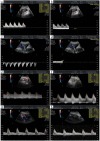
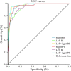
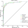
The objective was to assess the sensitivities and accuracies of Doppler ultrasound parameters in the second and third trimester of hypertensive pregnancies in determining perinatal outcomes. 1,054 pregnancies were retrospectively categorized into three groups (healthy pregnancies (HP, n = 988), pregnancies of hypertensive women (HypP, n = 30) and high-risk hypertension pregnancies (HRHypP, n = 36), depending on gestational hypertension as well as fetal birth weights and pregnancy outcomes. Systolic/diastolic ratio (S/D), resistance index (RI), pulsatility index (PI) of the bilateral uterine artery, umbilical artery and vein as well as venous flow velocity data were monitored by Doppler ultrasound. At 20-27 and 28-32 gestational weeks, uterine artery PIs and RIs were significantly higher in the HRHypP group than in the HP and HypP patients. At gestational weeks 20-27 and 28-32 left plus right PI data with cut-off values of 2.35 and 1.73 indicated a risk of stillbirth, premature pregnancy termination and a birth weight of less than 2,500 g with sensitivities of 94.4% and 93.1% as well as specificities of 95.2% and 90.1%, respectively.
Figures


References
Publication types
MeSH terms
LinkOut - more resources
Full Text Sources
Other Literature Sources
Medical
Research Materials
Miscellaneous

