. 2017 Jan 16:7:40573.
doi: 10.1038/srep40573.

Efficient generation of hPSC-derived midbrain dopaminergic neurons in a fully defined, scalable, 3D biomaterial platform

Affiliations

Efficient generation of hPSC-derived midbrain dopaminergic neurons in a fully defined, scalable, 3D biomaterial platform

Maroof M Adil et al. Sci Rep. .

Abstract

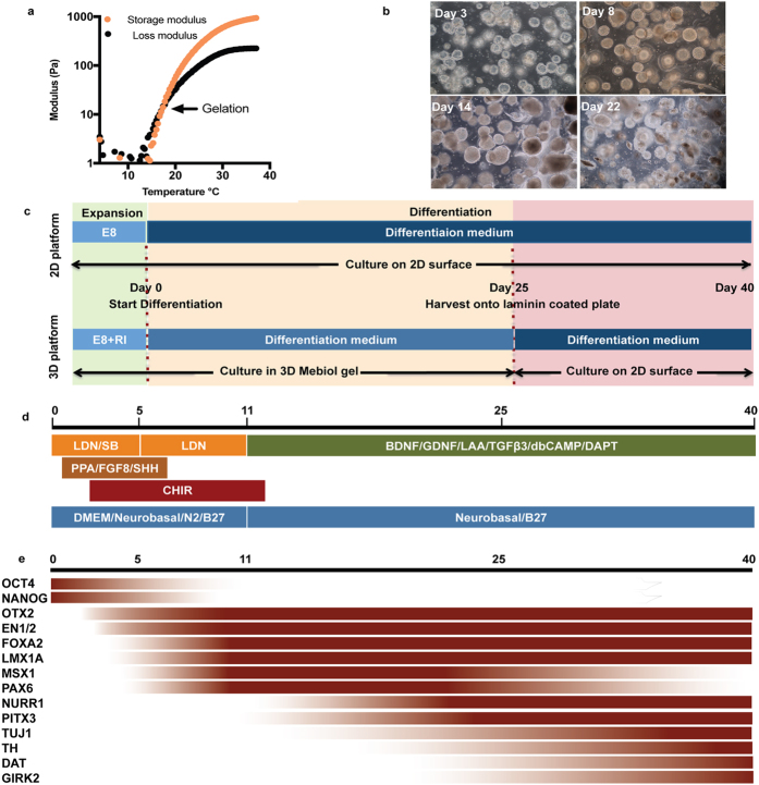
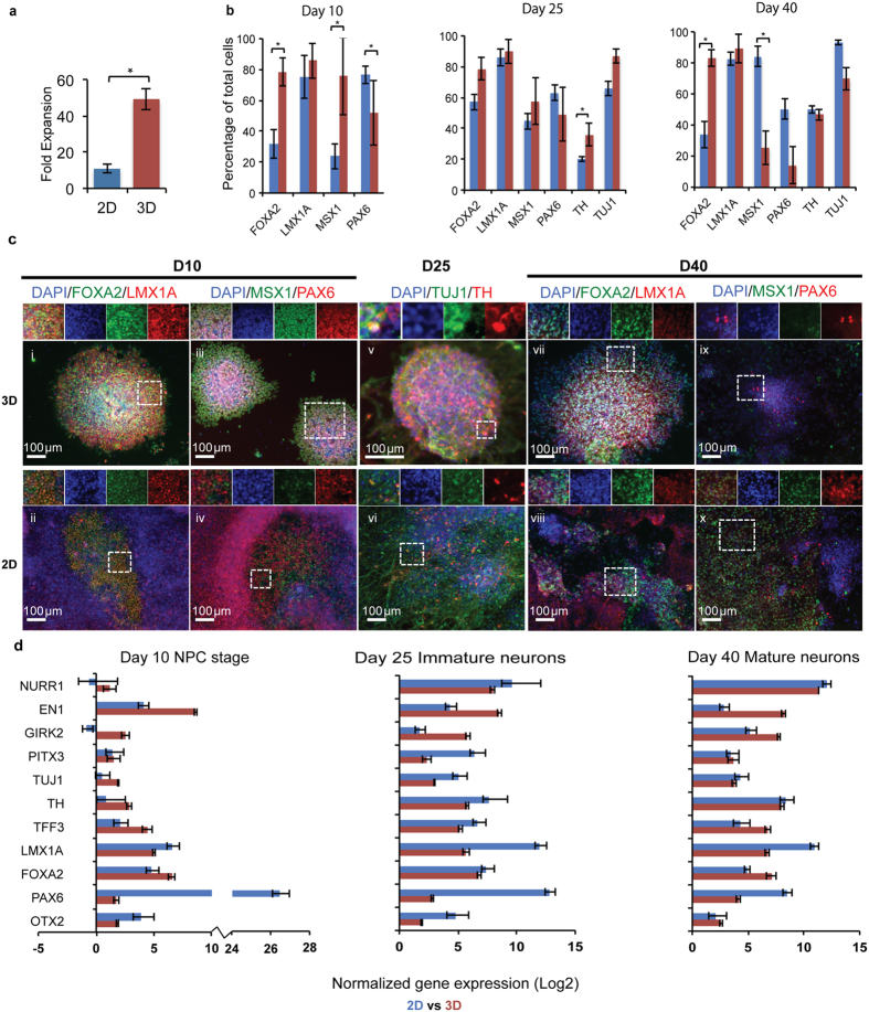
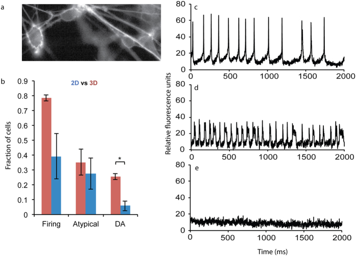
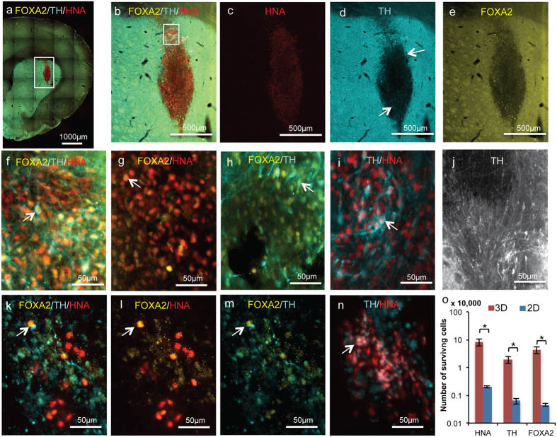
Pluripotent stem cells (PSCs) have major potential as an unlimited source of functional cells for many biomedical applications; however, the development of cell manufacturing systems to enable this promise faces many challenges. For example, there have been major recent advances in the generation of midbrain dopaminergic (mDA) neurons from stem cells for Parkinson's Disease (PD) therapy; however, production of these cells typically involves undefined components and difficult to scale 2D culture formats. Here, we used a fully defined, 3D, thermoresponsive biomaterial platform to rapidly generate large numbers of action-potential firing mDA neurons after 25 days of differentiation (~40% tyrosine hydroxylase (TH) positive, maturing into 25% cells exhibiting mDA neuron-like spiking behavior). Importantly, mDA neurons generated in 3D exhibited a 30-fold increase in viability upon implantation into rat striatum compared to neurons generated on 2D, consistent with the elevated expression of survival markers FOXA2 and EN1 in 3D. A defined, scalable, and resource-efficient cell culture platform can thus rapidly generate high quality differentiated cells, both neurons and potentially other cell types, with strong potential to accelerate both basic and translational research.


Figures

Figure 1
Figure 1. Material properties of 10 w/v % PNIPAAM-PEG are amenable for maintaining pluripotency for hPSCs and generation of hESC derived neurons.
(a) Rheological measurements of storage and loss moduli of PNIPAAm-PEG gels demonstrating the thermoresponsive liquid to solid transition. Traces are representative of 3 independent experiments. (b) Brightfield images of H1 hESC-derived clusters in PNIPAAm-PEG 3D platform at different stages during the mDA differentiation process. Images are representative of n = 4 independent experiments. (c) Schematic showing differentiation conditions for 2D and 3D culture. (d) Diagram for mDA differentiation protocol. (e) Pictorial representation of anticipated expression levels of different markers of interest during mDA neuronal development, based on previously reported trends.
Figure 2
Figure 2. Comparative characterization of H1 hESC derived mDA neurons generated on 2D versus 3D platforms.
(a) Fold expansion of mDA neurons after 25 days of differentiation in 3D vs. 2D culture. Data are presented as mean ± s.e.m. from n = 3 independent experiments. *p < 0.05 for Student’s t test. (b) Quantitative immunocytochemistry comparing mDA marker expression at Days 10, 25, and 40 between 2D (blue) and 3D (red) cultures. Data are presented as mean ± s.e.m. for n = 3 independent experiments. *p < 0.05 for Student’s t test. (c) Representative fluorescence images highlighting significant differences between 2D and 3D cultures, corresponding to data presented in (b); (i–ii, vii–viii) FOXA2 (green)/LMX1A (red) and (iii–iv, ix–x) MSX1 (green)/PAX6 (red) at Days 10 and 40, and (v–vi) TH (red)/TUJ1 (green) at Day 25. Nuclei are labeled with DAPI (blue). A region of interest (dashed white square) is highlighted along with individual channels for each marker above each image. Scale bars, 100 μm. (d) Comparative gene expression analysis at Days 10, 25, and 40 between 2D (blue) and 3D (red) generated mDA neurons. Data are presented as the mean ± standard deviation from triplicates.
Figure 3
Figure 3. Electrophysiological properties of H1 hESC-derived mDA neurons.
(a) Representative image of voltage sensitive dye labeled mDA neuron culture. (b) Comparative quantification of the fraction of total cell population, from neurons generated on 2D (blue) or in 3D (red) platforms, firing distinct action potentials. Data are presented as mean ± s.e.m from n = 3 independent experiments, for images from 42 total cells in 3D and 48 total cells in 2D. *p < 0.05 for Student’s t test. Representative fluorescence intensity traces corresponding to mDA neuron action potential firing (c), atypical firing (d), and non-specific noise (e).
Figure 4
Figure 4. In vivo survival of 3D or 2D platform generated mDA neurons in rats.
(a–e) Graft morphology at 6 weeks post-implantation for mDA neurons generated in 3D, showing expression of HNA, TH, and FOXA2. (f) Inset from (b), showing coexpression of TH and FOXA2 in surviving HNA+ cells. White arrow shows an example of a cell coexpressing HNA, FOXA2 and TH. Coexpression of FOXA2 and HNA (g), of TH and FOXA2 (h), and of TH and HNA (i), with white arrow showing examples of each. (j) TH+ neurite growth within the graft core. (k–n) Graft at 6 weeks post-implantation for mDA neurons generated on 2D, showing expression of HNA, TH, and FOXA2. (k) Infrequent coexpression of FOXA2 and TH in HNA+ surviving cells (k) of FOXA2 and HNA (l), and of FOXA2 and TH (m), shown by white arrows. (n) Coexpression of TH and HNA, shown by white arrow. (o) Quantification of total number of HNA+, TH+, and FOXA2+ surviving cells from 4 animals/group for mDA neurons generated in 3D (red bars) or in 2D (blue bars). Data are presented as mean ± s.e.m.
Figure 5
Figure 5. Maturation and synaptic connections formed in 3D generated cell grafts and 6 weeks post-implantation in rats.
(a–e) Representative image showing coexpression of STEM121 (red), TH (green), and human synaptophysin (hSyn, blue). (f–j) Representative image showing STEM121 positive human cells (red) expressing human synaptophysin (hSyn, blue) at the interface with DARPP32+ striatal neurons (green). Images are representative of 4 animals.

References

    1. Kriks S. et al. Dopamine neurons derived from human ES cells efficiently engraft in animal models of Parkinson’s disease. Nature 480, 547–551 (2011). - PMC - PubMed
    1. Grealish S. et al. Human ESC-derived dopamine neurons show similar preclinical efficacy and potency to fetal neurons when grafted in a rat model of Parkinson’s Disease. Cell Stem Cell 15, 653–665 (2014). - PMC - PubMed
    1. Doi D. et al. Isolation of human induced pluripotent stem cell-derived dopaminergic progenitors by cell sorting for successful transplantation. Stem Cell Reports 2, 337–350 (2014). - PMC - PubMed
    1. Kirkeby A. et al. Generation of regionally specified neural progenitors and functional neurons from human embryonic stem cells under defined conditions. Cell Rep. 1, 703–714 (2012). - PubMed
    1. Lindvall O. Dopaminergic neurons for Parkinson’s therapy. Nat. Biotechnol. 30, 56–8 (2012). - PubMed

Publication types

MeSH terms