Review
. 2017 Mar;255(3):435-447.
doi: 10.1007/s00417-016-3580-y. Epub 2017 Jan 14.

Adaptation, perceptual learning, and plasticity of brain functions

Affiliations
Review

Adaptation, perceptual learning, and plasticity of brain functions

Jonathan C Horton et al. Graefes Arch Clin Exp Ophthalmol. 2017 Mar.

Abstract

The capacity for functional restitution after brain damage is quite different in the sensory and motor systems. This series of presentations highlights the potential for adaptation, plasticity, and perceptual learning from an interdisciplinary perspective. The chances for restitution in the primary visual cortex are limited. Some patterns of visual field loss and recovery after stroke are common, whereas others are impossible, which can be explained by the arrangement and plasticity of the cortical map. On the other hand, compensatory mechanisms are effective, can occur spontaneously, and can be enhanced by training. In contrast to the human visual system, the motor system is highly flexible. This is based on special relationships between perception and action and between cognition and action. In addition, the healthy adult brain can learn new functions, e.g. increasing resolution above the retinal one. The significance of these studies for rehabilitation after brain damage will be discussed.

Keywords: Adaptation; Brain plasticity; Motor cortex; Perceptual learning; Rehabilitation; Visual cortex.


Conflict of interest statement

Conflict of interest

All authors certify that they have no affiliations with or involvement in any organization or entity with any financial interest (such as honoraria; educational grants; participation in speakers’ bureaus; membership, employment, consultancies, stock ownership, or other equity interest; and expert testimony or patent-licensing arrangements), or non-financial interest (such as personal or professional relationships, affiliations, knowledge or beliefs) in the subject matter or materials discussed in this manuscript.

Ethical statement

For this type of study formal consent is not required.

Figures

Fig. 1
Fig. 1
CT scan showing an acute left parietal hematoma, causing a right homonymous hemianopia. A CT scan performed 5 months later shows damage to the left optic radiations. The visual field cut never recovered
Fig. 2
Fig. 2
Retinal input is conveyed to the primary (striate) cortex by a two-neuron chain, crossing a single relay in the lateral geniculate nucleus. Injury at any point cuts off visual perception, although a small projection (green shading) from the lateral geniculate to area MT allows “blindsight” in patients with homonymous hemianopia caused by a post-chiasmal lesion (pink shading). After Polyak (1957)
Fig. 3
Fig. 3
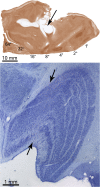
(Top) Flattened tissue section reacted for cytochrome oxidase showing a large lesion (arrow) of the primary visual cortex in a monkey. (Bottom) The lesion produced a swath of cell loss, visible in a Nissl-stained section, running through all layers of the lateral geniculate nucleus (arrows). Relay neurons in the lateral geniculate die because their axon terminals are destroyed in the cortex
Fig. 4
Fig. 4
a Right occipital lobe, with red shading to indicate the primary visual cortex. A large stroke (blue shading) from occlusion of the posterior cerebral artery is shown. b The calcarine fissure is opened to reveal the primary visual cortex. The stroke extends even beyond the edge of the semi-flattened cortex, except posteriorly, where cortex is supplied by the middle cerebral artery. c Flattened sheet of cortex, marking the boundaries of the stroke in (b) with a dashed line. Months after stroke, some recovery may occur at the fringes of the infarct, reducing the amount of cortical damage (shown schematically by shrinkage of the blue shading). However, the stroke still extends far beyond the borders of the primary visual cortex, so no recovery of visual field along the vertical meridian should be expected
Fig. 5
Fig. 5
Fixational eye movements during fixation of a cross are asymmetric towards the blind side, shown for right hemianopia: a assessment by scanning laser ophthalmoscope (SLO), example of one patient. b in conventional perimetry (schematic), the visual field defect and the blind spot are shifted towards the blind side. c distribution of fixational eye movements in 25 patients with right hemianopia with absent or small (<4°) macular sparing assessed by SLO (based on 1000 video fields per patient): the mean is shifted to 2.6 degrees to the right and is significantly different from normal distribution modified after [12]
Fig. 6
Fig. 6
Exploration of a natural scene in right hemianopia: a) without eye movements, b) with scanning eye movements the field of gaze is utilized and obstacles, here the baby stroller, can be seen in time
Fig. 7
Fig. 7
Reading in hemianopia depends on the configuration of the field defect and the available perceptual span during one fixation: a In macular splitting, half of the reading visual field is covered and functionless, resulting in severe reading impairment. b In macular sparing, the reading visual field can be spared and reading can be normal. c A paracentral homonymous scotoma leads to severe reading impairment. d Eccentric fixation shifts the field defect towards the hemianopic side and creates a small perceptual strip along the vertical field border, a favorable adaptive mechanism
Fig. 8
Fig. 8
Improvement of visual acuity (preferential looking upper left, and VEP upper right), and visual field size (bottom; isopters shown for years) during early years of life. Results of preferential looking and visual evoked potentials from [47]

References

    1. Sincich LC, Adams DL, Economides JR, Horton JC. Transmission of spike trains at the retinogeniculate synapse. J Neurosci. 2007;27:2683–2692. doi: 10.1523/JNEUROSCI.5077-06.2007. - DOI - PMC - PubMed
    1. Gilbert CD, Wiesel TN. Receptive field dynamics in adult primary visual cortex. Nature. 1992;356:150–152. doi: 10.1038/356150a0. - DOI - PubMed
    1. Chino YM, Kaas JH, Smith EL, III, et al. Rapid reorganization of cortical maps in adult cats following restricted deafferentation in retina. Vision Res. 1992;32:789–796. doi: 10.1016/0042-6989(92)90021-A. - DOI - PubMed
    1. Horton JC, Hocking DR. Monocular core zones and binocular border strips in primate striate cortex revealed by the contrasting effects of enucleation, eyelid suture, and retinal laser lesions on cytochrome oxidase activity. J Neurosci. 1998;18(14):5433–5455. - PMC - PubMed
    1. Smirnakis SM, Brewer AA, Schmid MC, et al. Lack of long-term cortical reorganization after macaque retinal lesions. Nature. 2005;435(7040):300–307. doi: 10.1038/nature03495. - DOI - PubMed