. 2017 Mar;20(3):427-437.
doi: 10.1038/nn.4479. Epub 2017 Jan 16.

REM sleep selectively prunes and maintains new synapses in development and learning

Affiliations

REM sleep selectively prunes and maintains new synapses in development and learning

Wei Li et al. Nat Neurosci. 2017 Mar.

Abstract

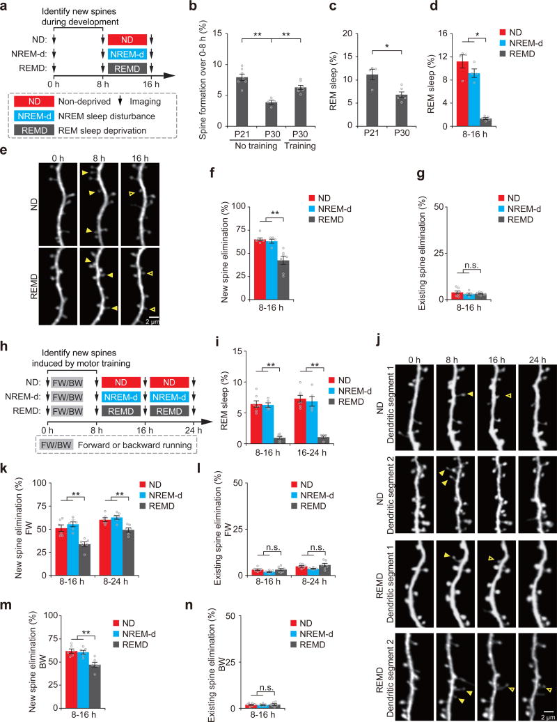
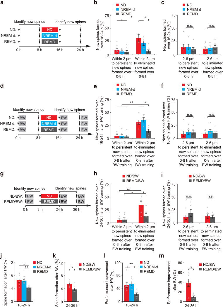
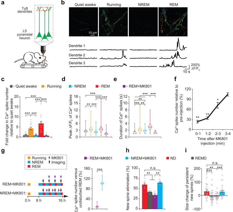
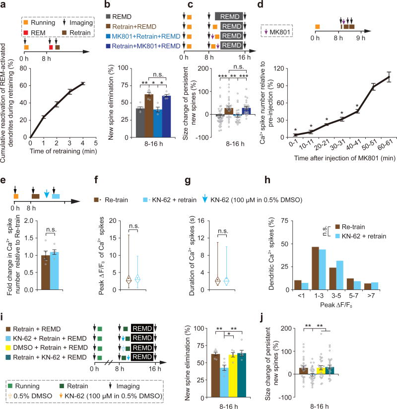
The functions and underlying mechanisms of rapid eye movement (REM) sleep remain unclear. Here we show that REM sleep prunes newly formed postsynaptic dendritic spines of layer 5 pyramidal neurons in the mouse motor cortex during development and motor learning. This REM sleep-dependent elimination of new spines facilitates subsequent spine formation during development and when a new motor task is learned, indicating a role for REM sleep in pruning to balance the number of new spines formed over time. Moreover, REM sleep also strengthens and maintains newly formed spines, which are critical for neuronal circuit development and behavioral improvement after learning. We further show that dendritic calcium spikes arising during REM sleep are important for pruning and strengthening new spines. Together, these findings indicate that REM sleep has multifaceted functions in brain development, learning and memory consolidation by selectively eliminating and maintaining newly formed synapses via dendritic calcium spike-dependent mechanisms.


Figures

Figure 1
Figure 1. REM sleep prunes newly formed spines during development and after learning
(a) Schematic of experimental design. After identification of new spines formed between hours 0–8, young mice at P21 were either left undisturbed (nondeprived control, ND), subjected to NREM sleep disturbance (NREM-d) or REM sleep deprivation (REMD) and new spines were imaged over the next 8 h. (b) The rate of new spine formation was significantly reduced as animals matured (P = 0.008; n = 7 and 4 mice at P21 and P30, respectively). Motor training significantly increased the formation of new spines in P30 mice; P = 0.008; n = 7 and 4 mice with and without motor training, respectively). (c) The amount of REM sleep was significantly lower in P30 mice than in P21 mice (P = 0.033, n = 6 P30 mice and 4 P21 mice). (d) The amount of REM sleep was significantly reduced in REMD mice as compared to ND and NREM-d mice between hours 8–16 (ND vs. REMD, P = 0.014; NREM-d vs. REMD, P = 0.014; n = 4, 4 and 5 ND, NREM-d and REMD mice, respectively). (e) Repeated imaging of dendritic spines on apical tuft dendrites of L5 pyramidal neurons in ND and REMD mice at P21. Filled arrowheads indicate new spines formed during hours 0–8. Some new spines (open arrowheads) persisted for the next 8 h. (f) The elimination rate of new spines (formed between hours 0–8) was significantly higher in ND or NREM-d mice than in REMD mice over hours 8–16 (ND vs. REMD, P = 0.002; NREM-d vs. REMD, P = 0.006; n = 7, 5 and 7 ND, NREM-d and REMD mice, respectively). (g) REMD did not affect the elimination of existing spines over hours 8–16 (n = 7, 5 and 7 ND, NREM-d and REMD mice, respectively). (h) Schematic of experimental design. After identification of motor-training-induced (forward running, FW; backward running, BW) new spines formed between hours 0–8, 1-month-old (P30) mice were either left undisturbed (ND), subjected to NREM sleep disturbance (NREM-d) or REM sleep deprivation (REMD) and new spines were imaged over the next 8–16 h. (i) The amount of REM sleep was significantly reduced in REMD mice as compared to ND and NREM-d mice between hours 8–16 (ND vs. REMD, P = 0.003; NREM-d vs. REMD, P = 0.006) and 16–24 (ND vs. REMD, P = 0.003; NREM-d vs. REMD, P = 0.006; n = 7, 5 and 6 ND, NREM-d and REMD mice, respectively). (j) Repeated imaging of dendritic spines before and 24 h after rotarod training in ND and REMD mice. Filled arrowheads indicate new spines formed during hours 0–8 after training. Some new spines (open arrowheads) persisted over the next 8–16 h. (k) The elimination rate of new spines (formed between 0–8 h after FW training) was significantly higher in ND or NREM-d mice than in REMD mice over the next 8–16 h (P = 0.004 for ND or NREM-d versus REMD over hours 8–16; P = 0.008 for ND or NREM-d versus REMD over hours 8–24; n = 6 mice for each group). (l) REMD did not affect the elimination of existing spines between hours 8–24 (over hours 8–16: ND vs. REMD, P = 0.748; NREM-d vs. REMD, P = 0.261; over hours 8–24: ND vs. REMD, P = 0.749; NREM-d vs. REMD, P = 0.078; n = 6 mice for each group). (m) The elimination rate of new spines formed 0–8 h after backward-running (BW) training was significantly higher in ND or NREM-d mice than in REMD mice over the next 8 h (P = 0.006 for ND or NREM-d versus REMD, n = 6 mice for each group). (n) REMD did not affect the elimination rate of existing spines over hours 8–16 (n = 6 mice for each group). Data are presented as mean ± s.e.m. Each point in b–d,f,g,i,k–n represents data from one animal. *P < 0.05, **P < 0.01, n.s. = not significant. Scale bars, 2 μm.
Figure 2
Figure 2. REM sleep-dependent spine elimination facilitates subsequent new spine formation at nearby sites
(a) Experimental design for examining the relationship between new spines formed during hours 0–8 and new spines formed over hours 16–24. New spines were identified in P21 mice over hours 0–8 and classified as persistent or eliminated based on their fate over hours 8–16. (b) New spines formed over hours 16–24 were rarely located within 2 μm of persistent new spines formed over hours 0–8. Significantly larger percentages of new spines formed over hours 16–24 were located within 2 μm of transient new spines formed over hours 0–8 in ND and NREM-d mice than in REMD mice (P = 0.01 for ND or NREM-d versus REMD, n = 7, 5 and 7 mice for ND, NREM-d and REMD, respectively). (c) The percentages of new spines formed over hours 16–24 and located 2–6 μm away from persistent or eliminated new spines formed over hours 0–8 were comparable among ND, NREM-d and REMD mice (n = 7, 5 and 7 mice for ND, NREM-d and REMD, respectively). (d) Experimental design for examining the relationship between the elimination of new spines induced by BW and the formation of new spines induced by FW. New spines were identified in mice subjected to BW between hours 0–8. Eight hours after ND, NREM-d or REMD sleep, the animals were subjected to FW. (e) FW-induced new spines were rarely located within 2 μm of persistent BW-induced new spines. Significantly larger percentages of FW-induced new spines were located within 2 μm of transient BW-induced new spines in ND and NREM-d mice than in REMD mice (P = 0.01 for ND or NREM-d versus REMD, n = 6 mice for each group). (f) The percentages of new spines formed over hours 16–24 after FW and located 2–6 μm from persistent or eliminated new spines formed over hours 0–8 after BW were comparable among ND, NREM-d and REMD mice (n = 6 mice for each group). (g–i) Experimental design as in d–f but for examining the relationship between the elimination of new spines induced by FW and the formation of new spines induced by BW; similar results were achieved. (j) New spines were induced by FW between hours 16–24 at a significantly higher rate in ND and NREM-d mice than in REMD mice (ND vs. REMD, P = 0.01; NREM-d vs. REMD, P = 0.004; n = 6 mice for each group). (k) New spines were induced by BW between hours 24–36 at a significantly higher rate in ND mice than in REMD mice (P = 0.037, n = 6 mice for each group). (l) The rotarod performance improvement of FW was significantly smaller in REMD mice compared to in ND and NREM-d mice over hours 16–24 (ND vs. REMD, P = 0.004; NREM-d vs. REMD, P = 0.007; n = 7, 7 and 6 ND, NREM-d and REMD mice, respectively). (m) The rotarod performance improvement of BW was significantly lower in REMD mice compared to ND mice over hours 24–36 (P = 0.016, n = 6 mice for each group). Data are presented as mean ± s.e.m. *P < 0.05, **P < 0.01, n.s. = not significant.
Figure 3
Figure 3. REM sleep strengthens a fraction of new spines formed during development and learning
(a) Schematic of experimental design. After identification of new spines formed between hours 0–8, young mice P21 were left either undisturbed (ND), subjected to NREM sleep disturbance (NREM-d) or REM sleep deprivation (REMD), and new spines were imaged over the next 8 h. (b) Repeated imaging of dendritic spines in ND and REMD mice at P21. Arrowheads indicate new spines that formed during hours 0–8 and persisted over time. (c) The average size of persistent new spines in P21 mice increased over time in ND (32 persistent new spines from 7 mice over hours 8–16) and NREM-d mice (24 persistent new spines from 5 mice), as compared to REMD mice (55 persistent new spines from 7 mice) (ND vs. REMD, P = 0.006; NREM-d vs. REMD, P = 0.018). (d) REMD mice did not show substantial differences in the size of existing spines compared to ND and NREM-d mice at P21 (72, 63 and 91 randomly selected existing spines from 7, 5 and 7 ND, NREM-d and REMD mice, respectively). (e) Schematic of experimental design. After identification of motor-training-induced new spines formed between hours 0–8, P30 mice were left either undisturbed (ND), subjected to NREM sleep disturbance (NREM-d) or REM sleep deprivation (REMD), and new spines were imaged over the next 8–16 h. (f) Repeated imaging of dendritic spines before and 24 h after motor training in ND and REMD mice. Arrowheads indicate new spines that formed 0–8 h after training and persisted over time. (g) The average size of persistent new spines induced by FW increased over time in ND (22 new spines from 6 mice over hours 8–16, and 54 new spines from 16 mice over hours 8–24) and NREM-d mice (37 spines from 7 mice over hours 8–16, and 31 spines from 7 mice over hours 8–24) but not in REMD mice (37 spines from 6 mice over hours 8–16, and 75 spines from 16 mice over hours 8–24) (over hours 8–16: ND vs. REMD, P = 0.001; NREM-d vs. REMD, P = 0.0001; over hours 8–24: ND vs. REMD, P = 1.19 × 10⁻¹⁰; NREM-d vs. REMD, P = 4.52 × 10⁻⁸). (h) REMD mice did not show any effects on the size of existing spines as compared to ND or NREM-d mice at P30 (45, 59 and 40 randomly selected existing spines for ND, NREM-d and REMD mice, respectively). (i) The average size of persistent new spines induced by BW increased over time in ND (23 new spines from 6 mice over hours 8–16) and NREM-d mice (23 spines from 6 mice over hours 8–16) but not in REMD mice (57 spines from 6 mice over hours 8–16) (ND vs. REMD, P = 4.25 × 10⁻⁴; NREM-d vs. REMD, P = 8.28 × 10⁻⁴). (j) REMD mice did not show any effects on the size of existing spines as compared to ND or NREM-d mice (45, 48 and 56 randomly selected existing spines for ND, NREM-d and REMD mice, respectively). (k) The rotarod performance improvement of FW running was significantly lower in REMD mice compared to that in ND and NREM-d mice over hours 0–24 (ND vs. REMD, P = 0.032; NREM-d vs. REMD, P = 0.046; n = 7, 7 and 6 ND, NREM-d and REMD mice, respectively). Data are presented as mean ± s.e.m. *P < 0.05, **P < 0.01, ***P < 0.001, n.s. = not significant. Scale bars, 2 μm.
Figure 4
Figure 4. REM sleep facilitates long-term maintenance of new spines during development and learning
(a) Experimental design for examining the long-term survival of new spines formed over hours 0–8 in P21 mice. After undisturbed sleep (ND), NREM-d or REMD for 8 h, new spines identified over hours 0–8 were examined over 4 d. The overall survival rate of new spines formed over hours 0–8 was significantly lower in REMD mice than in ND and NREM-d mice over 4 d (16 h: ND vs. REMD, P = 0.002; NREM-d vs. REMD, P = 0.006; n = 7, 5 and 7 ND, NREM-d and REMD mice, respectively; 4 d: ND vs. REMD, P = 0.013;NREM-d vs. REMD, P = 0.008; n = 4, 5 and 5 ND, NREM-d and REMD mice, respectively). (b) After 8 h of REM sleep deprivation, a significantly larger percentage of new spines formed between hours 0–8 were eliminated in REMD mice compared to in ND mice or NREM-d mice over 4 d (ND vs. REMD, P = 0.013;NREM-d vs. REMD, P = 0.008; n = 4, 5 and 5 ND, NREM-d and REMD mice, respectively). (c) New spines persisted over 4 d were strengthened in size between hours 8–16 in ND and NREM-d control mice, whereas new spines eliminated over 4 d were not strengthened during the same period (21 persistent and 22 eliminated new spines from 9 mice). (d) New spines persisted or eliminated over 4 d were not strengthened in size over hours 8–16 in REMD mice (2 persistent and 35 eliminated new spines from 5 mice). (e) Experimental design for examining the long-term survival of FW-induced new spines. New spines were identified in P30 mice subjected to FW on an accelerated rotarod over hours 0–8. After undisturbed sleep or REMD for 16 h, the animals were subjected to BW or no training, and the survival of the new spines identified over hours 0–8 were examined between hours 24–36. The overall survival rate of new spines formed after FW (hours 0–8) was significantly lower between hours 8–36 in REMD mice than in ND mice after BW (ND + BW vs. REMD + BW, P = 0.02; REMD without BW vs. REMD + BW, P = 0.011; n = 4, 6, 4 and 6 ND without BW, ND + BW, REMD without BW and REMD + BW mice, respectively). (f) After BW training, a significantly larger percentage of new spines formed over hours 0–8 were eliminated between hours 24–36 in REMD mice compared to in ND mice or nontrained mice (ND + BW vs. REMD + BW, P = 0.004; REMD without BW vs. REMD + BW, P = 0.009; n = 4, 6, 4 and 6 ND without BW, ND + BW, REMD without BW and REMD + BW mice, respectively). (g) Persistent new spines but not eliminated new spines over hours 24–36 were strengthened in size between hours 8–24 in ND mice (29 persistent and 10 eliminated new spines from 10 mice). (h) New spines persistent or eliminated over hours 24–36 were not strengthened in size between hours 8–24 in REMD mice (24 persistent and 23 eliminated new spines from 10 mice). Data are presented as mean ± s.e.m. *P < 0.05, **P < 0.01, n.s. = not significant.
Figure 5
Figure 5. Dendritic Ca2+ spikes occurring during REM sleep are important for new spine elimination and strengthening
(a) Two-photon Ca2+ imaging of apical tuft dendrites of L5 pyramidal neurons in head-restrained mice on a treadmill during quiet awake, running, NREM sleep and REM sleep. (b) Ca2+ imaging of apical tuft dendrites under various states. Ca2+ fluorescence traces of three dendrites over 1 min are shown. Images at 3 timepoints with a 10-s interval are represented by three different colors. Scale bar, 10 μm. (c,d) The number and peak amplitude of dendritic Ca2+ spikes during REM sleep were comparable to those during running but significantly larger than those during either NREM sleep, quiet awake state or REM sleep with MK801 application (for number: P = 1.82 × 10⁻⁵, 0.082, 9.37 × 10⁻⁵ and 1.66 × 10⁻⁴ for REM sleep vs. quiet awake, running, NREM sleep and REM sleep + MK801, respectively; n = 13, 12, 9, 13 and 8 quiet awake, running, NREM, REM and REM + MK801 mice, respectively; for peak amplitude: P = 2.82 × 10⁻⁴, 0.373, 9.09 × 10⁻⁸ and 1.77 × 10⁻¹² for REM sleep vs. quiet awake, running, NREM sleep and REM sleep + MK801, respectively; n = 89, 124, 88, 274 and 47 spikes for quiet awake, running, NREM, REM and REM + MK801, respectively). (e) The durations of dendritic Ca2+ spikes during REM sleep were significantly larger than those during other states (P = 3.01 × 10⁻¹⁷, 0.0007, 1.88 × 10⁻¹⁰ and 1.47 × 10⁻¹³ for REM sleep vs. quiet awake, running, NREM sleep and REM sleep + MK801, respectively). (f) Brief injection of MK801 (3 pulses, 50 ms each) into the primary motor cortex blocked dendritic Ca2+ spikes during quiet awake over the next 2–3 min (n = 10 mice). (g) More than 90% of dendritic Ca2+ spikes during REM sleep were blocked after pulsed injection of MK801 at the beginning of each REM sleep episode but not during NREM sleep (n = 46 episodes of REM sleep with MK801 injection from 4 mice; 51 episodes of NREM sleep with MK801 injection from 4 mice). (h) Injections of MK801 during REM sleep but not during NREM sleep, reduced the elimination rate of learning-induced new spines (ND vs. REM + MK801, P = 0.006; ND vs. NREM + MK801, P = 0.297; REM + MK801 vs. NREM + MK801, P = 0.006; n = 6, 5, 5 and 6 mice for ND, REMD, REM + MK801 and NREM + MK801, respectively). (i) Injection of MK801 during REM sleep, but not during NREM sleep, reduced the size increase of persistent new spines formed after treadmill training (ND vs. REM + MK801, P = 0.0019; ND vs. NREM + MK801, P = 0.801; REM + MK801 vs. NREM + MK801, P = 0.0005; n = 25, 40, 26 and 29 new spines from 6, 5, 5 and 6 mice for ND, REMD, REM + MK801 and NREM + MK80, respectively). Data are presented as mean ± s.e.m. In d, e and g, box and whisker plots show the means (central dot), medians (central line in the box), ranges between 25th and 75th percentiles (box) and minimum–maximum range (whiskers). Each point in c and h represents data from one animal. **P < 0.01, ***P < 0.001, n.s. = not significant.
Figure 6
Figure 6. Motor training-induced Ca2+ spikes prune and strengthen new spines in REMD mice
(a) Majority of dendrites exhibiting Ca2+ spikes during 1-min REM sleep also showed Ca2+ spikes during 4-min retraining (n = 4 mice). (b) 40-min retraining led to increased elimination of new spines in REMD mice. Injection of MK801 before retraining, but not after retraining, blocked retraining-induced elimination of new spines (n = 5, 6, 4 and 5 mice for REMD, retraining + REMD, MK801 + retraining + REMD, and retraining + MK801 + REMD, respectively). (c) 40-min retraining led to the potentiation of persistent new spines in REMD mice. Injection of MK801 before retraining, but not after retraining, blocked retraining-induced strengthening of persistent new spines (40, 26, 25 and 25 new spines from 5, 6, 4 and 5 mice for REMD, retraining + REMD, MK801 + retraining + REMD, and retraining + MK801 + REMD, respectively). (d) The number of retraining-induced dendritic Ca2+ spikes was reduced substantially for ~40 min after repeated injections of MK801 (10 pulses) at the beginning of retraining (n = 6 mice). (e–h) The number, average peak amplitude, duration and peak amplitude distribution of dendritic calcium spikes over a period of 1 min were not affected by local application of KN-62 (n = 5 mice). (i) Application of KN-62 before retraining, but not after retraining, blocked retraining-induced elimination of new spines in REMD mice (retraining vs. KN-62 + retraining, P = 0.008; KN-62 + retraining vs. retraining + KN-62, P = 0.006; n = 6, 6, 5 and 6 mice for retraining, KN62 + retraining, DMSO + retraining and retraining + KN62, respectively). (j) Application of KN-62 before retraining, but not after retraining, blocked retraining-induced strengthening of persistent new spines (retraining vs. KN-62 + retraining, P = 0.007; KN-62 + retraining vs. retraining + KN-62, P = 0.003; n = 26, 31, 25 and 26 new spines from 6, 6, 5 and 6 mice for retraining, KN62 + retraining, DMSO + retraining and retraining + KN62, respectively). In f and g, box and whisker plots show means (central dot), medians (central line in the box), ranges between 25th and 75th percentiles (box) and minimum–maximum ranges (whiskers). Data are presented as mean ± s.e.m. Each point in b, e and i represents data from one animal. *P < 0.05, **P < 0.01, ***P < 0.001, n.s. = not significant.

References

    1. Roffwarg HP, Muzio JN, Dement WC. Ontogenetic development of the human sleep-dream cycle. Science. 1966;152:604–619. doi: 10.1126/science.152.3722.604. - DOI - PubMed
    1. Jouvet M. Paradoxical sleep and the nature-nurture controversy. Prog Brain Res. 1980;53:331–346. doi: 10.1016/S0079-6123(08)60073-4. - DOI - PubMed
    1. Siegel JM. The REM sleep-memory consolidation hypothesis. Science. 2001;294:1058–1063. doi: 10.1126/science.1063049. - DOI - PMC - PubMed
    1. Hobson JA. REM sleep and dreaming: towards a theory of protoconsciousness. Nat Rev Neurosci. 2009;10:803–813. doi: 10.1038/nrn2716. - DOI - PubMed
    1. Karni A, Tanne D, Rubenstein BS, Askenasy JJ, Sagi D. Dependence on REM sleep of overnight improvement of a perceptual skill. Science. 1994;265:679–682. - PubMed