. 2017 Jan 16;17(1):44.
doi: 10.1186/s12903-017-0341-1.

In vitro effects of hyaluronic acid on human periodontal ligament cells

Affiliations

In vitro effects of hyaluronic acid on human periodontal ligament cells

Masako Fujioka-Kobayashi et al. BMC Oral Health. .

Abstract

Background: Hyaluronic acid (HA) has been reported to have a positive effect on periodontal wound healing following nonsurgical and surgical therapy. However, to date, a few basic in vitro studies have been reported to investigating the potential of HA on human periodontal ligament (PDL) cell regeneration. Therefore, the aim of this study was to investigate the effect of HA on PDL cell compatibility, proliferation, and differentiation in vitro.

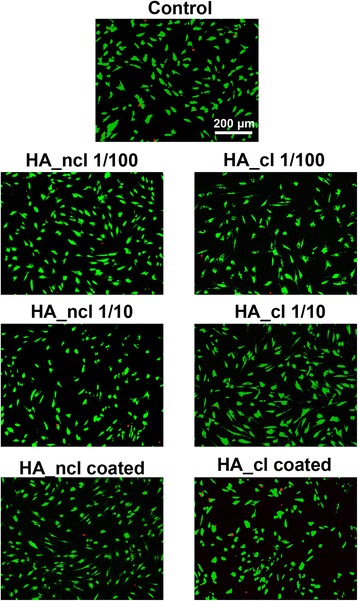
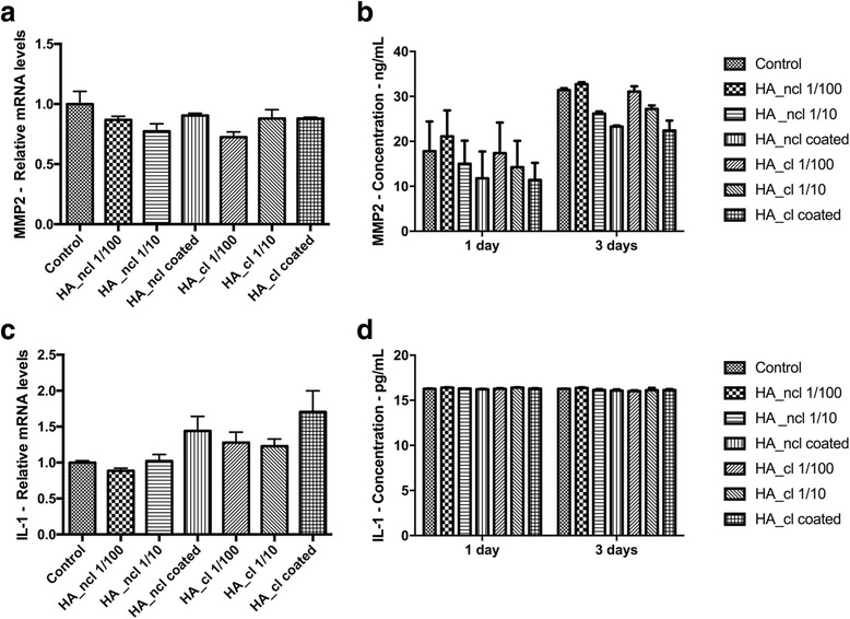
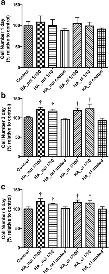
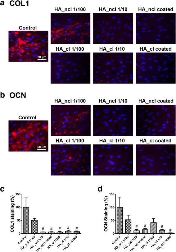
Methods: Either non-cross-linked (HA_ncl) or cross-linked (HA_cl) HA was investigated. Human PDL cells were seeded in 7 conditions as follows (1) Control tissue culture plastic (TCP) (2) dilution of HA_ncl (1:100), (3) dilution of HA_ncl (1:10), 4) HA_ncl directly coated onto TCP, (5) dilution of HA_cl (1:100), 6) dilution of HA_cl (1:10) and (7) HA_cl directly coated onto TCP. Samples were then investigated for cell viability using a live/dead assay, an inflammatory reaction using real-time PCR and ELISA for MMP2, IL-1 and cell proliferation via an MTS assay. Furthermore, the osteogenic potential of PDL cells was assessed by alkaline phosphatase(ALP) activity, collagen1(COL1) and osteocalcin(OCN) immunostaining, alizarin red staining, and real-time PCR for genes encoding Runx2, COL1, ALP, and OCN.

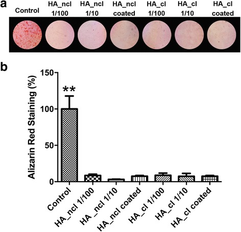
Results: Both HA_ncl and HA_cl showed high PDL cell viability (greater than 90%) irrespective of the culturing conditions. Furthermore, no significant difference in both mRNA and protein levels of proinflammatory cytokines, including MMP2 and IL-1 expression was observed. Both diluted HA_ncl and HA_cl significantly increased cell numbers compared to the controlled TCP samples at 3 and 5 days. HA_ncl and HA_cl in standard cell growth media significantly decreased ALP staining, COL1 immunostaining and down-regulated early osteogenic differentiation, including Runx2, COL1, and OCN mRNA levels when compared to control samples. When osteogenic differentiation medium (ODM) was added, interestingly, the expression of early osteogenic markers increased by demonstrating higher levels of COL1 and ALP expression; especially in HA 1:10 diluted condition. Late stage osteogenic markers remained inhibited.

Conclusions: Both non-cross-linked and cross-linked HA maintained high PDL cell viability, increased proliferation, and early osteogenic differentiation. However, HA was consistently associated with a significant decrease in late osteogenic differentiation of primary human PDL cells. Future in vitro and animal research is necessary to further characterize the effect of HA on periodontal regeneration.

Keywords: Connective tissue regeneration; Hyaluronan; Hyaluronic acid; Periodontal regeneration; Soft tissue regeneration.


Figures

Fig. 1
Fig. 1
Cell viability staining of primary human primary PDL cells exposed to control (TCP), non-cross-linked HA (HA_ncl) and cross-linked HA (HA_cl) surfaces. For cell viability, Live-Dead staining was done with viable cell appearing in green and dead cells in red. The results from these experiments demonstrated that both HA_ncl and HA_cl are highly biocompatible at dilutions of 1:100 and 1:10 as well as pre-coated onto cell culture plastic
Fig. 2
Fig. 2
Real-time PCR of PDL cells seeded with HA_ncl and HA_cl for genes encoding (a) matrix metalloproteinase-2 (MMP2), (c) Interleukin-1 (IL-1), at 1 days post seeding. Protein release at 1 and 3 days of (b) MMP2, (d) IL-1, (** denotes significantly higher than all other modalities among HA treated groups, p < 0.05)
Fig. 3
Fig. 3
Proliferation assay of PDL cells seeded with HA_ncl and HA_cl at (a) 1, (b) 3 and (c) 5 days post seeding. It was found that both HA and HA_cl at dilutions of 1:100 and 1:10 significantly increased cell numbers at 3 days and 5 days post seeding when compared to control samples († denotes significantly higher than control, p < 0.05)
Fig. 4
Fig. 4
Real-time PCR of PDL cells seeded with HA_ncl and HA_cl treatment for genes encoding (a, b) Runx2, (c, d) Collagen 1 alpha 2 (COL1a2), (e, f) alkaline phosphatase (ALP) and (g, h) osteocalcin (OCN) at 7 days post seeding. Cells were treated (a, c, e, g) in regular growth medium or (b, d, f, h) with ODM. (* denotes significant difference, p < 0.05; # denotes significantly lower than control, p < 0.05; † denotes significantly higher than control, p < 0.05; ** denotes significantly higher than all other treatment modalities, p < 0.05)
Fig. 5
Fig. 5
Alkaline phosphatase staining of PDL cells treated by HA_ncl and HA_cl (a) in growth medium or (b) within ODM at 7 days post seeding. Both HA_ncl and HA_cl significantly decreased ALP staining without ODM while with ODM both HA_ncl and HA_cl significantly increased ALP staining when compared to control samples (* denotes significant difference, p < 0.05; # denotes significantly lower than control, p < 0.05; † denotes significantly higher than control, p < 0.05; ** denotes significantly higher than all other treatment modalities, p < 0.05)
Fig. 6
Fig. 6
Immunofluorescent COL1 staining and OCN staining at 14 days post cell seeding with HA_ncl and HA_cl. (a) The merged images of immunofluorescent detection of COL1 (red) and DAPI (blue). (b) The merged images of immunofluorescent detection of OCN (red) and DAPI (blue). (c, d) Quantified data of (c) COL1 and (d) OCN immunostaining at 14 days (# denotes significantly lower than control, p < 0.05)
Fig. 7
Fig. 7
Alizarin red staining denoting mineralization at 14 days post seeding. (a) Alizarin red staining images and (b) quantified data of alizarin red staining from colour thresholding software for PDL cells treated with HA_ncl or HA_cl (** denotes significantly higher than all other treatment modalities, p < 0.05). It was found that both HA_ncl and HA_cl treatment significantly decreased alizarin red staining

Similar articles

Cited by

References

    1. Toole BP. Hyaluronan in morphogenesis. Semin Cell Dev Biol. 2001;12(2):79–87. doi: 10.1006/scdb.2000.0244. - DOI - PubMed
    1. Bertolami CN, Messadi DV. The role of proteoglycans in hard and soft tissue repair. Crit Rev Oral Biol Med. 1994;5(3–4):311–337. doi: 10.1177/10454411940050030601. - DOI - PubMed
    1. Pilloni A, Rimondini L, De Luca M, Bernard GW. Effect of hyaluronan on calcification-nodule formation from human periodontal ligament cell culture. J Appl Biomater Biomech. 2003;1(1):84–90. - PubMed
    1. Chen WY, Abatangelo G. Functions of hyaluronan in wound repair. Wound Repair Regen. 1999;7(2):79–89. doi: 10.1046/j.1524-475X.1999.00079.x. - DOI - PubMed
    1. Donegan GC, Hunt JA, Rhodes N. Investigating the importance of flow when utilizing hyaluronan scaffolds for tissue engineering. J Tissue Eng Regen Med. 2010;4(2):83–95. doi: 10.1002/term.208. - DOI - PubMed

MeSH terms