. 2017 Feb 1;10(2):151-162.
doi: 10.1242/dmm.025593. Epub 2017 Jan 12.

A model of type 2 diabetes in the guinea pig using sequential diet-induced glucose intolerance and streptozotocin treatment

Affiliations

A model of type 2 diabetes in the guinea pig using sequential diet-induced glucose intolerance and streptozotocin treatment

Brendan K Podell et al. Dis Model Mech. .

Abstract

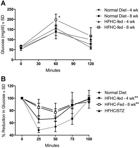
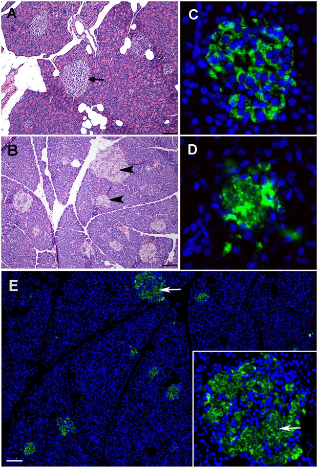
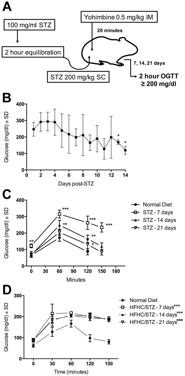
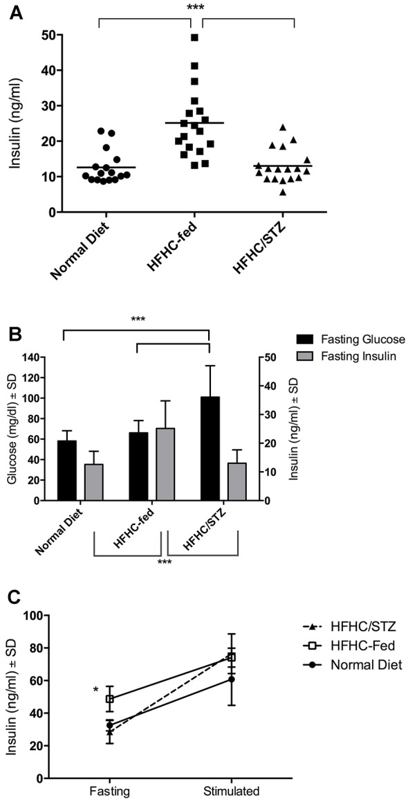
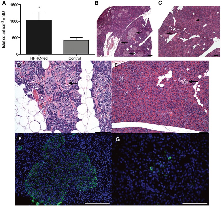
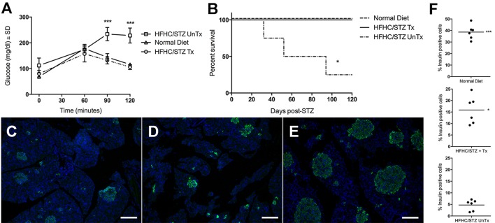
Type 2 diabetes is a leading cause of morbidity and mortality among noncommunicable diseases, and additional animal models that more closely replicate the pathogenesis of human type 2 diabetes are needed. The goal of this study was to develop a model of type 2 diabetes in guinea pigs, in which diet-induced glucose intolerance precedes β-cell cytotoxicity, two processes that are crucial to the development of human type 2 diabetes. Guinea pigs developed impaired glucose tolerance after 8 weeks of feeding on a high-fat, high-carbohydrate diet, as determined by oral glucose challenge. Diet-induced glucose intolerance was accompanied by β-cell hyperplasia, compensatory hyperinsulinemia, and dyslipidemia with hepatocellular steatosis. Streptozotocin (STZ) treatment alone was ineffective at inducing diabetic hyperglycemia in guinea pigs, which failed to develop sustained glucose intolerance or fasting hyperglycemia and returned to euglycemia within 21 days after treatment. However, when high-fat, high-carbohydrate diet-fed guinea pigs were treated with STZ, glucose intolerance and fasting hyperglycemia persisted beyond 21 days post-STZ treatment. Guinea pigs with diet-induced glucose intolerance subsequently treated with STZ demonstrated an insulin-secretory capacity consistent with insulin-independent diabetes. This insulin-independent state was confirmed by response to oral antihyperglycemic drugs, metformin and glipizide, which resolved glucose intolerance and extended survival compared with guinea pigs with uncontrolled diabetes. In this study, we have developed a model of sequential glucose intolerance and β-cell loss, through high-fat, high-carbohydrate diet and extensive optimization of STZ treatment in the guinea pig, which closely resembles human type 2 diabetes. This model will prove useful in the study of insulin-independent diabetes pathogenesis with or without comorbidities, where the guinea pig serves as a relevant model species.

Keywords: Animal model; Glucose intolerance; Guinea pig; Insulin-independent diabetes; Streptozotocin; Type 2 diabetes.


Conflict of interest statement

Competing interests

The authors declare no competing or financial interests.

Figures

Fig. 1.
Fig. 1.
Guinea pigs fed HFHC diet develop impaired glucose tolerance and suppressed response to insulin. Fasted or nonfasted guinea pigs were challenged with an oral glucose bolus or exogenous insulin, respectively, and blood glucose was measured over time. (A) Response to an oral glucose challenge in guinea pigs that consumed HFHC diet for 4 or 8 weeks (n=20), compared with normal-diet controls (n=20). Fasting hyperglycemia is absent, but delayed glucose clearance is evident 60 min after glucose challenge in HFHC-fed guinea pigs. *P<0.05, two-way ANOVA. (B) Response of HFHC-fed guinea pigs to an insulin tolerance test (n=5) after 4 or 8 weeks of HFHC feeding or in HFHC/STZ guinea pigs compared with normal-diet controls (n=5). Impaired tissue sensitivity to insulin is apparent in HFHC-fed and HFHC/STZ guinea pigs, based on failure to reduce blood glucose over time compared with normal-diet controls. **P<0.01, based on mean area under the curve, Student's t-test.
Fig. 2.
Fig. 2.
Optimized STZ treatment leads to specific cytotoxicity of insulin-producing cells. Images are representative of guinea pigs developing hyperglycemia after treatment with an optimized SC dose of anomer-equilibrated STZ at 200 mg/kg administered after an intramuscular injection of yohimbine. (A) Pancreatic islets from a nondiabetic, normal-diet control guinea pig (arrow) demonstrating histological morphology in the absence of diabetogenic treatment. (B) Morphological changes 48 h after STZ treatment in pancreas of a guinea pig with confirmed hyperglycemia that was fed a normal diet. Cell death has occurred in the majority of islet cells (arrowheads), while a minority of the cells remain viable. (C) Immunofluorescent detection of pro-insulin (green) in pancreatic islets in a normal-diet control guinea pig indicates that the majority of islet cells are insulin-producing β cells (blue, Hoechst nuclear counterstain). (D,E) Immunofluorescent detection of pro-insulin in a guinea pig 48 h after receiving an optimized dose of STZ. Disruption of cellular and nuclear morphology is uniform across all islets and is specific to insulin-expressing β cells (arrows and inset). Scale bar: 100 µm.
Fig. 3.
Fig. 3.
Development of stable STZ-induced diabetes requires coexisting diet-induced impaired glucose tolerance. (A) Diagrammatic representation of optimized STZ treatment to induce hyperglycemia in guinea pigs fed either normal or HFHC diet. STZ powder is dissolved in citrate buffer at 100 mg/ml, then incubated for 2 h to allow for α/β anomer equilibration. Twenty minutes before STZ treatment, guinea pigs are administered a 0.5 mg/kg dose of the α2 agonist yohimbine, by the intramuscular (IM) route. Guinea pigs are then given a single 200 mg/kg subcutaneous (SC) injection of anomer-equilibrated STZ. Diabetic hyperglycemia is determined by an oral glucose tolerance test (OGTT) at day 7, 14 and 21, based on a 2 h blood glucose ≥200 mg/dl. (B) Blood glucose concentrations were measured daily at random in nonfasted, STZ-treated guinea pigs fed a normal diet (n=5). Hyperglycemia, observable 24 h after STZ treatment, has a trend of steady decline over the course of 14 days. *P<0.05 compared with day 1 post-STZ, one-way ANOVA. (C) Glucose tolerance was evaluated in STZ-treated guinea pigs (n=5) fed a normal diet by standardized oral glucose challenge on day 7, 14 and 21 after STZ treatment, and compared with normal-diet controls (n=5). Fasting hyperglycemia and diabetic glucose intolerance is evident 7 days after STZ treatment, but glucose intolerance diminishes over time, returning to nondiabetic tolerance after 21 days. **P<0.01, ***P<0.001, compared with mean glucose of normal-diet controls at 0, 60 and 120 min, or day 21 challenge of STZ guinea pigs at 150 min, two-way ANOVA. (D) HFHC-fed guinea pigs were treated with STZ after 8 weeks of consuming HFHC diet. Response to an oral glucose challenge is documented over 7, 14 and 21 days after STZ treatment that was preceded by 8 weeks of HFHC feeding (n=5 or 10). STZ treatment results in stable fasting hyperglycemia and diabetic glucose intolerance persisting to 21 days after treatment when guinea pigs are challenged in the presence of HFHC diet-induced impaired glucose tolerance. *P<0.05, ***P<0.001, compared with mean glucose of normal-diet controls at that measurement time point, one-way ANOVA based on mean area under the curve.
Fig. 4.
Fig. 4.
Compensatory hyperinsulinemia maintains euglycemia in HFHC-fed guinea pigs but is lost with STZ treatment. (A) Insulin was detected by direct ELISA in serum from fasted guinea pigs fed a normal diet (n=16), HFHC diet (n=18) or HFHC diet and treated with STZ (HFHC/STZ, n=18). Fasting hyperinsulinemia is present in guinea pigs after 8 weeks of consuming the HFHC diet. Treatment of HFHC-fed guinea pigs with STZ leads to impairment of the compensatory insulin response; however, insulin production is retained in HFHC/STZ guinea pigs at a level comparable to normal-diet controls. ***P<0.001, one-way ANOVA. (B) Fasting blood glucose concentrations were measured from fasting samples taken simultaneously for insulin quantification by direct ELISA. Fasting hyperglycemia is absent in the presence of compensatory hyperinsulinemia in HFHC-fed guinea pigs. Fasting hyperglycemia is present in HFHC/STZ guinea pigs after impairment of the compensatory insulin response. ***P<0.001, one-way ANOVA. (C) Glucose-stimulated insulin secretion performed in guinea pigs after 8 weeks of HFHC feeding, then repeated 21 days after receiving STZ. Residual function is retained in β cells after STZ treatment (n=3). *P<0.05, paired one-way ANOVA.
Fig. 5.
Fig. 5.
Morphological alterations in pancreatic islets demonstrate a compensatory response to HFHC diet and STZ-induced β-cell loss. (A) Pancreas tissue area was quantified using morphometry software and frequency of islets expressed per centimeter squared of total pancreatic tissue (n=5 per group). HFHC-fed guinea pigs develop islet hyperplasia after 8 weeks of consuming HFHC diet, consistent with insulin resistance and a compensatory response. *P<0.05, Student's t-test. (B) Histological morphology of pancreas from an HFHC-fed guinea pig. Islets are both enlarged and more frequent compared with normal-diet controls (arrows). (C) Pancreas from a normal-diet control guinea pig demonstrates the typical frequency and size of pancreatic islets in the absence of diabetogenic treatment (arrows). Scale bar: 100 µm. (D) Representative islet morphology from a guinea pig fed HFHC diet for an extended period of 6 months. Degenerative changes are present in enlarged islets, indicated by deposition of fibrous connective tissue (arrow). Scale bar: 100 µm. (E) Islet morphology 3 weeks after STZ treatment in an HFHC/STZ guinea pig. Islets are reduced in size after treatment with STZ (arrow). Scale bar: 200 µm. (F) Immunofluorescent detection of pro-insulin (green) in an enlarged islet from an HFHC-fed guinea pig after 8 weeks of consuming the diet. Enlarged islets contain a high frequency of insulin-producing β cells. Blue is Hoechst nuclear counterstain. Scale bar: 100 µm. (G) Immunofluorescent detection of pro-insulin in an islet from an HFHC/STZ guinea pig 3 weeks after STZ treatment. There is an overall reduction in the frequency of insulin-producing β cells 3 weeks after STZ treatment. Scale bar: 100 µm.
Fig. 6.
Fig. 6.
HFHC/STZ guinea pigs develop insulin-independent diabetes that is responsive to oral antihyperglycemic therapy. (A) Diabetic HFHC/STZ guinea pigs received either a combination of metformin and glipizide therapy (HFHC/STZ Tx, n=4), initiated 21 days after STZ treatment, or remained with uncontrolled diabetes (HFHC/STZ UnTx, n=4). Responses to an oral glucose challenge were compared with normal-diet controls (n=2). Metformin and glipizide treatment in HFHC/STZ guinea pigs reverses diabetic glucose intolerance and fasting hyperglycemia. ***P<0.001, one-way ANOVA. (B) Survival was tracked for 120 days in HFHC/STZ guinea pigs that were either treated with metformin and glipizide or remained with uncontrolled diabetes, compared with normal-diet control guinea pigs. Antihyperglycemic therapy significantly improves survival in guinea pigs with HFHC/STZ diabetes. *P<0.05. (C) Immunofluorescent detection of pro-insulin after 120 days of treatment in HFHC/STZ guinea pigs treated with metformin and glipizide combination therapy. Reduced β-cell frequency remains present after 120 days in the presence of combination therapy. (D) Immunofluorescent detection of pro-insulin in HFHC/STZ guinea pigs with uncontrolled diabetes. Insulin production appears similar to those receiving combination therapy. (E) Immunofluorescent detection of pro-insulin in a normal-diet control guinea pig from the survival group, demonstrating a greater frequency of β cells in the absence of diabetogenic treatment. (F) Quantitation of pro-insulin-positive cells expressed as a percentage of tissue area from six representative fields per treatment group. HFHC/STZ treatment reduces total β-cell mass, which increases in guinea pigs treated with metformin and glipizide. *P<0.05, ***P<0.001, compared with untreated HFHC/STZ guinea pigs, one-way ANOVA. Scale bars: 100 μm.

Similar articles

Cited by

References

    1. Aomine M., Nobe S. and Arita M. (1990). The making of diabetic guinea pigs by streptozotocin and high incidence of triggered activity in the ventricular muscle. Jpn. J. Physiol. 40, 651-663. 10.2170/jjphysiol.40.651 - DOI - PubMed
    1. Ar'Rajab A. and Ahrén B. (1991). Effects of yohimbine and nicotinic acid on insulin secretion in islet transplanted streptozotocin-diabetic rats. Diabetes Res. Clin. Pract. 11, 81-87. 10.1016/0168-8227(91)90095-U - DOI - PubMed
    1. Aslan M., Ozcan F. and Kucuksayan E. (2013). Increased small dense LDL and decreased paraoxonase enzyme activity reveals formation of an atherogenic risk in streptozotocin-induced diabetic guinea pigs. J. Diabetes Res. 2013, 860190 10.1155/2013/860190 - DOI - PMC - PubMed
    1. Beagley J., Guariguata L., Weil C. and Motala A. A. (2014). Global estimates of undiagnosed diabetes in adults. Diabetes Res. Clin. Pract. 103, 150-160. 10.1016/j.diabres.2013.11.001 - DOI - PubMed
    1. Bell R. H. Jr. and Hye R. J. (1983). Animal models of diabetes mellitus: physiology and pathology. J. Surg. Res. 35, 433-460. 10.1016/0022-4804(83)90034-3 - DOI - PubMed

Publication types

MeSH terms