Review
. 2017 Mar 5;372(1715):20160157.
doi: 10.1098/rstb.2016.0157.

Multiple shared mechanisms for homeostatic plasticity in rodent somatosensory and visual cortex

Affiliations
Review

Multiple shared mechanisms for homeostatic plasticity in rodent somatosensory and visual cortex

Melanie A Gainey et al. Philos Trans R Soc Lond B Biol Sci. .

Abstract

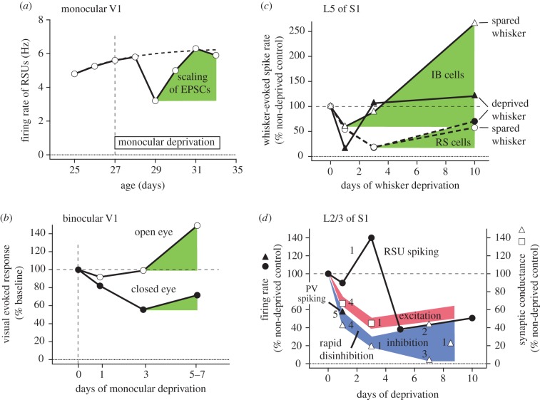
We compare the circuit and cellular mechanisms for homeostatic plasticity that have been discovered in rodent somatosensory (S1) and visual (V1) cortex. Both areas use similar mechanisms to restore mean firing rate after sensory deprivation. Two time scales of homeostasis are evident, with distinct mechanisms. Slow homeostasis occurs over several days, and is mediated by homeostatic synaptic scaling in excitatory networks and, in some cases, homeostatic adjustment of pyramidal cell intrinsic excitability. Fast homeostasis occurs within less than 1 day, and is mediated by rapid disinhibition, implemented by activity-dependent plasticity in parvalbumin interneuron circuits. These processes interact with Hebbian synaptic plasticity to maintain cortical firing rates during learned adjustments in sensory representations.This article is part of the themed issue 'Integrating Hebbian and homeostatic plasticity'.

Keywords: firing rate homeostasis; homeostatic plasticity; inhibition; sensory cortex; somatosensory cortex; whisker.


Figures

Figure 1.
Figure 1.
Forms of firing rate homeostasis in V1 and S1 in vivo. (a) Mean firing rate of regular spiking units (RSU) in L2–L4 of monocular V1, following contralateral monocular deprivation (MD). MD initially reduces firing rates, which then return to normal despite continued deprivation. Dashed curve, schematic of normal firing rate development. Green, FRH attributed to synaptic scaling of mEPSCs. Data from Hengen et al. [8]. (b) Average population response in binocular V1 to open- and closed-eye visual stimuli following contralateral MD. MD rapidly decreases closed-eye visual responses, followed by a slower increase in both open- and closed-eye responses that is attributable to synaptic scaling (green). Points show average data from Frenkel & Bear [9] and Kaneko et al. [10]. (c) Effect of D-row whisker deprivation on whisker-evoked spiking in L5 IB and regular spiking (RS) pyramidal cells. In RS cells, deprivation reduces both deprived and spared whisker responses, followed by partial recovery. IB cells show a similar depression-recovery sequence, with preferential response gain for spared whisker responses. In both cell types, response recovery or potentiation are mediated by synaptic scaling (green). Data from Greenhill et al. [11]. (d) Rapid disinhibition during whisker deprivation in S1. Whisker-evoked spiking in L2/3 pyramidal cells is initially maintained and even transiently increased for 1–3 days following D-row whisker deprivation, prior to subsequent depression (circles). During this 1–3 day period, there is already a substantial weakening of excitatory synaptic drive to L2/3 neurons (red), evident as reduced whisker-evoked EPSCs in L2/3 neurons in vivo and reduced L4-evoked EPSCs in L2/3 pyramidal neurons in S1 slices (squares). Counteracting this loss of excitation is a rapid decrease in inhibition (blue), as reflected by reduced whisker-evoked IPSCs in L2/3 neurons in vivo and reduced L4-evoked and recurrent L2/3-evoked IPSCs in L2/3 pyramidal cells in S1 slices (open triangles). Rapid disinhibition also occurs in V1, as seen by reduced visual-evoked spiking of PV neurons following 1 day of MD (filled triangle). Data from 1: Li et al. [12]; 2: House et al. [13]; 3: Shao et al. [14]; 4: Gainey, SFN abstract [15]; 5: Kuhlman et al. [16].

References

    1. Hengen KB, Torrado Pacheco A, McGregor JN, Van Hooser SD, Turrigiano GG. 2016. Neuronal firing rate homeostasis is inhibited by sleep and promoted by wake. Cell 165, 180–191. (10.1016/j.cell.2016.01.046) - DOI - PMC - PubMed
    1. Turrigiano GG, Leslie KR, Desai NS, Rutherford LC, Nelson SB. 1998. Activity-dependent scaling of quantal amplitude in neocortical neurons. Nature 391, 892–896. (10.1038/36103) - DOI - PubMed
    1. Burrone J, O'Byrne M, Murthy VN. 2002. Multiple forms of synaptic plasticity triggered by selective suppression of activity in individual neurons. Nature 420, 414–418. (10.1038/nature01242) - DOI - PubMed
    1. Turrigiano G. 2012. Homeostatic synaptic plasticity: local and global mechanisms for stabilizing neuronal function. Cold Spring Harb. Perspect. Biol. 4, a005736 (10.1101/cshperspect.a005736) - DOI - PMC - PubMed
    1. Desai NS, Cudmore RH, Nelson SB, Turrigiano GG. 2002. Critical periods for experience-dependent synaptic scaling in visual cortex. Nat. Neurosci. 5, 783–789. (10.1038/nn878) - DOI - PubMed

Publication types