Review
. 2017 Jun;14(6):347-364.
doi: 10.1038/nrclinonc.2016.212. Epub 2017 Jan 17.

Beyond the margins: real-time detection of cancer using targeted fluorophores

Affiliations
Review

Beyond the margins: real-time detection of cancer using targeted fluorophores

Ray R Zhang et al. Nat Rev Clin Oncol. 2017 Jun.

Abstract

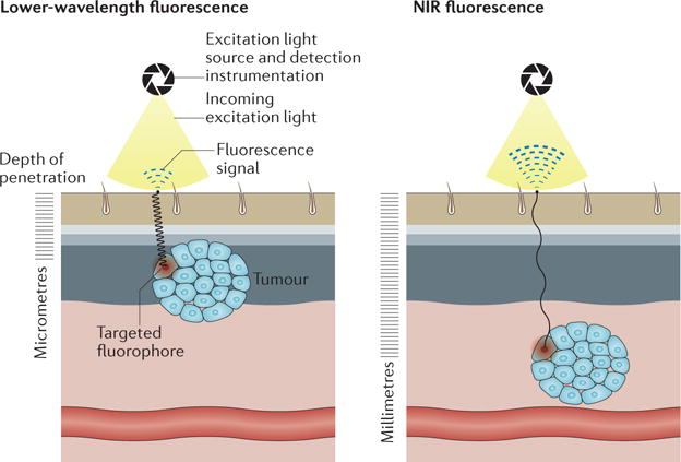
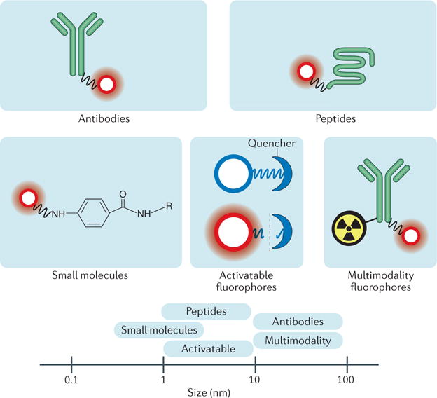
Over the past two decades, synergistic innovations in imaging technology have resulted in a revolution in which a range of biomedical applications are now benefiting from fluorescence imaging. Specifically, advances in fluorophore chemistry and imaging hardware, and the identification of targetable biomarkers have now positioned intraoperative fluorescence as a highly specific real-time detection modality for surgeons in oncology. In particular, the deeper tissue penetration and limited autofluorescence of near-infrared (NIR) fluorescence imaging improves the translational potential of this modality over visible-light fluorescence imaging. Rapid developments in fluorophores with improved characteristics, detection instrumentation, and targeting strategies led to the clinical testing in the early 2010s of the first targeted NIR fluorophores for intraoperative cancer detection. The foundations for the advances that underline this technology continue to be nurtured by the multidisciplinary collaboration of chemists, biologists, engineers, and clinicians. In this Review, we highlight the latest developments in NIR fluorophores, cancer-targeting strategies, and detection instrumentation for intraoperative cancer detection, and consider the unique challenges associated with their effective application in clinical settings.


Conflict of interest statement

Competing interests statement

J.J.G., A.N.P. and J.P.W. declare associations with Cellectar Biosciences. J.P.W. is the inventor of the APC analogues discussed in the Review. K.W.E is a consultant for the Bruker Corporation and is a co-founder of OnLume. R.R.Z., A.B.S., E.L.R., J.M.W. and J.S.K. declare no competing interests.

Figures

Figure 1
Figure 1. NIR fluorescence is more suitable for in vivo imaging applications than visible-light fluorescence
Near-infrared (NIR) fluorophores (700–900 nm) have deeper tissue penetration and lower background fluorescence than visible-light fluorescence, resulting in enhanced signal-to-noise ratios. The detection depths achievable with the currently available instrumentation ranges from millimeters with NIR fluorescence to micrometers with visible-range fluorescence.
Figure 2
Figure 2. Targeted fluorophores tested in clinical trials
Targeted fluorophores currently tested in clinical trials include small molecules, peptides, activatable fluorophores, antibodies, and multimodal fluorophores. Activatable fluorophores are initially quenched, but upon cleavage by enzymes, emit fluorescence. The relative sizes of these targeted fluorophores are displayed in the bottom panel.
Figure 3
Figure 3. Cetuximab-800CW fluorescence in a patient with EGFR-positive head and neck cancer
a | Preoperative bright-field image of a patient with a cutaneous squamous-cell carcinoma and b | preoperative fluorescence acquisition of the same tumour 3 days after the patient received intravenous cetuximab-800CW (50 mg) (NCT01987375). The image was acquired using an open-field fluorescence imaging device (Luna; Novadaq, Ontario, Canada). c | A bright-field image of the deep surface of a resected cutaneous squamous-cell carcinoma tumour from a different patient taken 3 days after cetuximab-800CW infusion (50 mg). d | Open-field fluorescence images (obtained with Luna, Novadaq) of the same tumour depicted in c shows areas of positive fluorescence. Additional resected specimen was taken from a deeper location than that of the primary specimen; positive areas of fluorescence were detected on e | the superficial surface, but not on f | the deep surface of the additional resection margin. g | Haematoxylin and eosin staining of 5 μm sectioned tissue from a primary tumour resected from another patient with a cutaneous squamous-cell carcinoma tumour taken 3 days after intravenous administration of cetuximab-800CW (50 mg). The outlined areas denote presence of cancer (assessed by a board-certified pathologist). h | Fluorescence scan (Odyssey; Li-COR Biosciences, Lincoln, Nebraska, USA) of the adjacent cut section, with outlined areas displaying bright fluorescence intensity.
Figure 4
Figure 4. Preclinical and clinical development of targeted fluorophores
The preclinical development phase entails target identification and validation, and fluorophore characterization. The clinical development of targeted fluorophores can require pairing with a device optimized for that particular fluorophore or ’combination product’. IDE, investigational device exemptions; IND, investigational new drug.

References

    1. Nguyen QT, Tsien RY. Fluorescence-guided surgery with live molecular navigation — a new cutting edge. Nat Rev Cancer. 2013;13:653–662. - PMC - PubMed
    1. Vahrmeijer AL, Hutteman M, van der Vorst JR, van de Velde CJ, Frangioni JV. Image-guided cancer surgery using near-infrared fluorescence. Nat Rev Clin Oncol. 2013;10:507–518. - PMC - PubMed
    1. Rosenthal EL, et al. Successful translation of fluorescence navigation during oncologic surgery: a consensus report. J Nucl Med. 2016;57:144–150. - PMC - PubMed
    1. Rosenthal EL, Warram JM, Bland KI, Zinn KR. The status of contemporary image-guided modalities in oncologic surgery. Ann Surg. 2015;261:46–55. - PMC - PubMed
    1. Yossepowitch O, et al. Positive surgical margins after radical prostatectomy: a systematic review and contemporary update. Eur Urol. 2014;65:303–313. - PubMed