. 2017 Jan-Feb;38(1):43-50.
doi: 10.1080/13816810.2016.1275018. Epub 2017 Jan 17.

Reduced penetrance in a large Caucasian pedigree with Stickler syndrome

Affiliations

Reduced penetrance in a large Caucasian pedigree with Stickler syndrome

Stuart W Tompson et al. Ophthalmic Genet. 2017 Jan-Feb.

Abstract

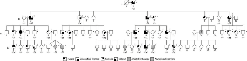
Background: In a four-generation Caucasian family variably diagnosed with autosomal dominant (AD) Stickler or Wagner disease, commercial gene screening failed to identify a mutation in COL2A1 or VCAN. We utilized linkage mapping and exome sequencing to identify the causal variant.

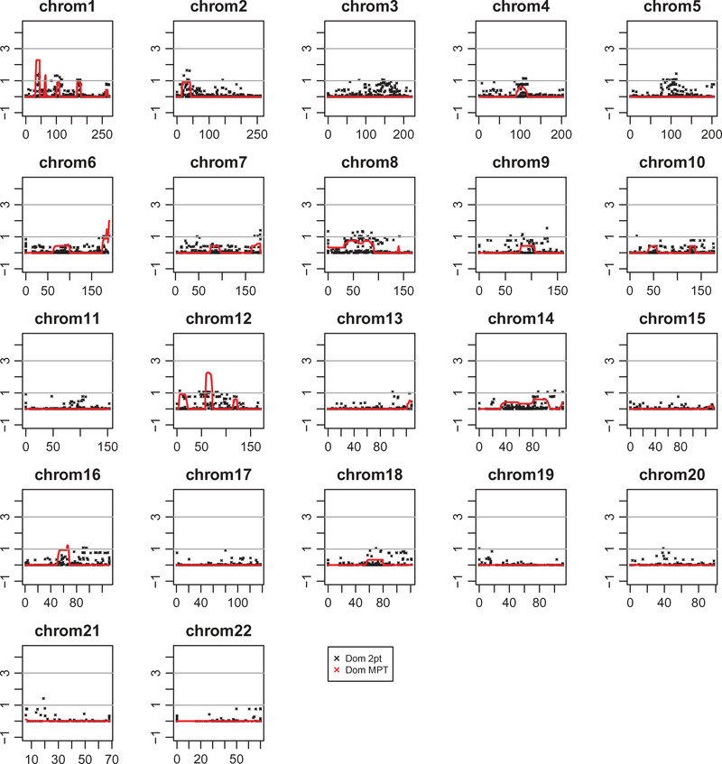
Materials and methods: Genomic DNA samples collected from 40 family members were analyzed. A whole-genome linkage scan was performed using Illumina HumanLinkage-24 BeadChip followed by two-point and multipoint linkage analyses using FASTLINK and MERLIN. Exome sequencing was performed on two affected individuals, followed by co-segregation analysis.

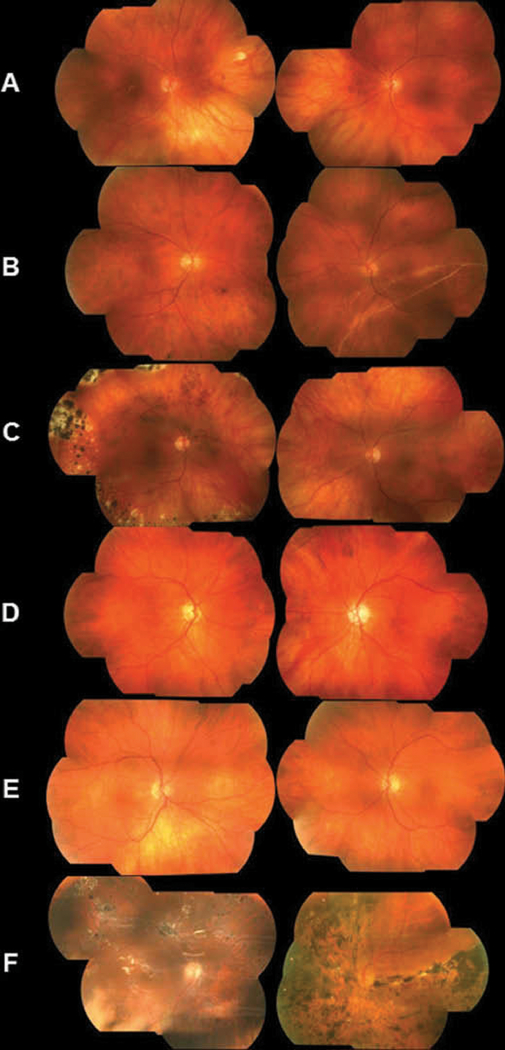
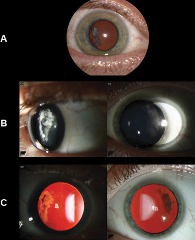
Results: Parametric multipoint linkage analysis using an AD inheritance model demonstrated HLOD scores > 2.00 at chromosomes 1p36.13-1p36.11 and 12q12-12q14.1. SIMWALK multipoint analysis replicated the peak in chromosome 12q (peak LOD = 1.975). FASTLINK two-point analysis highlighted several clustered chromosome 12q SNPs with HLOD > 1.0. Exome sequencing revealed a novel nonsense mutation (c.115C>T, p.Gln39*) in exon 2 of COL2A1 that is expected to result in nonsense-mediated decay of the RNA transcript. This mutation co-segregated with all clinically affected individuals and seven individuals who were clinically unaffected.

Conclusions: The utility of combining traditional linkage mapping and exome sequencing is highlighted to identify gene mutations in large families displaying a Mendelian inheritance of disease. Historically, nonsense mutations in exon 2 of COL2A1 have been reported to cause a fully penetrant ocular-only Stickler phenotype with few or no systemic manifestations. We report a novel nonsense mutation in exon 2 of COL2A1 that displays incomplete penetrance and/or variable age of onset with extraocular manifestations.

Keywords: Linkage; Stickler syndrome; Wagner syndrome; penetrance.


Conflict of interest statement

Declaration of interest

The authors report no conflicts of interest. The authors alone are responsible for the content and writing of this article.

Figures

Figure 1.
Figure 1.
Study family pedigree. The study family consisted of 70 individuals in four generations. Circles indicate females and squares indicate males. An individual’s affection status is depicted as black-shaded quadrants for the four main phenotypes (myopia, vitreoretinal changes, cataract, and scoliosis), according to the figure key shown. Gray-shaded shapes indicate clinically unaffected individuals who carried the mutation. The gray-shaded individual with a question mark (III:24) is known to be affected with Stickler syndrome by hearsay. * indicates DNA available for this study. ^ indicates samples used for whole-genome linkage analysis. # indicates samples were used for exome sequencing.
Figure 2.
Figure 2.
Fundus photos of affected individuals harboring the p.Gln39* mutation. (A) Photos of a 42-year-old female (III:3). The right eye underwent retinal detachment repair with scleral buckle at age 24. The vitreous was optically empty in both eyes. In the right eye, there were focal chorioretinal scars in the macula and mid-peripherally in the 2:00 meridian. Not visualized were inferior and temporal vitreous condensation and vitreous sheets bilaterally, as well as indentation from a scleral buckle in the right eye and chorioretinal scarring bilaterally. (B) Photos of the right eye of a male individual III:17 at age 43 (left image) and at age 46 (right image). At age 43, the right eye had an optically empty vitreous centrally with avascular sheets in the far temporal periphery. At age 46, the patient presented after one week of sudden visual field loss. A large retinal detachment could be observed, extending from the 12:00 to the 8:00 meridian, caused by a peripheral retinal hole at 02:00. (C) Photos of a 45-year-old female (III:21) showed peripheral scarring in the right eye from repair of a superior giant retinal tear occurring at the age of 15. The tear was treated with laser and a scleral buckle. Later, an inferior retinal hole was diagnosed and treated with laser and cryotherapy. Both eyes had an optically empty vitreous. (D) Photos of a 69-year-old male (II:2). The fundus exam was normal for the right eye. The left eye had an optically empty vitreous with avascular sheets in the periphery. Outside the extent of the left image was superior retinal scarring from the treatment of two retinal tears with pneumatic retinopexy. (E) Photos of a 66-year-old male (II:6) showed an optically empty vitreous in both eyes with avascular sheets in the far periphery. There was no central chorioretinal scarring, but the left eye had some scarring in the far superior periphery (not shown). (F) Photos of a 58-year-old male (II:11). The right eye sustained several retinal tears and detachments and was treated on multiple occasions with laser, cryotherapy, scleral buckle, vitrectomy, and finally placement of silicone oil. Multiple retinal scars were observed as well as light reflections from the oil. The left eye exhibited extensive chorioretinal scarring and pigmentation after treatment of the retinal detachment and scleral buckle placement.
Figure 3.
Figure 3.
Slit-lamp and retro-illumination photos of a father (III:18) and son (IV:17) with lamellar cataract in the temporal cortex of each eye. (A) Slit-lamp photo of the 42-year-old father’s right eye, which shows a temporal cortical cataract. The left eye underwent cataract extraction and intraocular lens placement for a visually significant cataract a year previously. (B) Slit-lamp photos of the 12-year-old son’s eyes, which show temporal cortical cataracts that are not visually significant. (C) Retro-illumination of the son’s eyes shows the distinctive temporal location of the cataracts.
Figure 4.
Figure 4.
Summary of two-point and multipoint linkage analyses using MERLIN. Plots of −log10 chromosome-wide significant p values (−log10P) for autosomal dominant parametric two-point and multipoint linkage analyses in 26 individuals. The maximum peak LOD scores were achieved on chromosomes 1p36.13–1p36.11 and 12q12–12q14.1. Y-axis represents −log10 ratio of the LOD score. X-axis represents chromosome position in centimorgans (cM).

Comment in

  • Type I membranous anomaly in Stickler syndrome.
    Alexander P, Poulson A, McNinch A, Richards A, Snead M. Alexander P, et al. Ophthalmic Genet. 2018 Jan-Feb;39(1):147. doi: 10.1080/13816810.2017.1326510. Epub 2017 May 30. Ophthalmic Genet. 2018. PMID: 28557656 No abstract available.

References

    1. Stickler GB, Belau PG, Farrell FJ, et al. Hereditary progressive arthro-ophthalmopathy. Mayo Clin Proc 1965;40:433–455. - PubMed
    1. Snead MP, McNinch AM, Poulson AV, et al. Stickler syndrome, ocular-only variants and a key diagnostic role for the ophthalmologist. Eye (Lond) 2011;25:1389–1400. - PMC - PubMed
    1. Snead MP, Yates JR. Clinical and molecular genetics of stickler syndrome. J Med Genet 1999;36:353–359. - PMC - PubMed
    1. Printzlau A, Andersen M. Pierre Robin sequence in Denmark: a retrospective population-based epidemiological study. Cleft Palate Craniofac J 2004;41:47–52. - PubMed
    1. Fertala A, Ala-Kokko L, Wiaderkiewicz R, Prockop DJ. Collagen II containing a Cys substitution for arg-alpha1–519. Homotrimeric monomers containing the mutation do not assemble into fibrils but alter the self-assembly of the normal protein. J Biol Chem 1997;272:6457–6464. - PubMed

MeSH terms

Supplementary concepts