Review
. 2017 Jul 1:176:195-206.
doi: 10.1016/j.physbeh.2017.01.027. Epub 2017 Jan 14.

Interoceptive modulation of neuroendocrine, emotional, and hypophagic responses to stress

Affiliations
Review

Interoceptive modulation of neuroendocrine, emotional, and hypophagic responses to stress

James W Maniscalco et al. Physiol Behav. .

Abstract

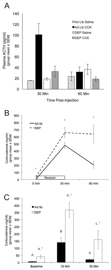
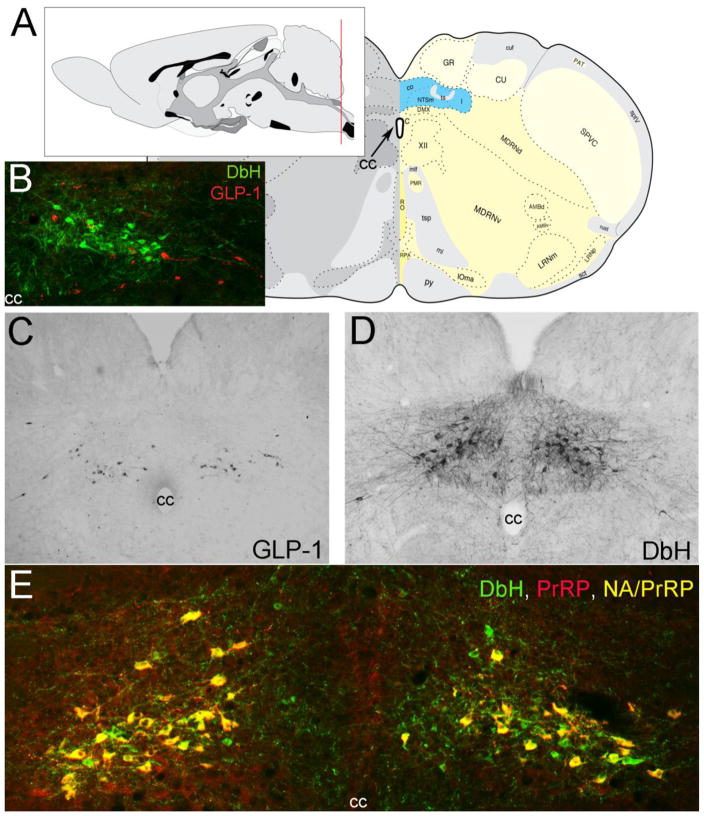
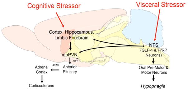
Periods of caloric deficit substantially attenuate many centrally mediated responses to acute stress, including neural drive to the hypothalamic-pituitary-adrenal (HPA) axis, anxiety-like behavior, and stress-induced suppression of food intake (i.e., stress hypophagia). It is posited that this stress response plasticity supports food foraging and promotes intake during periods of negative energy balance, even in the face of other internal or external threats, thereby increasing the likelihood that energy stores are repleted. The mechanisms by which caloric deficit alters central stress responses, however, remain unclear. The caudal brainstem contains two distinct populations of stress-recruited neurons [i.e., noradrenergic neurons of the A2 cell group that co-express prolactin-releasing peptide (PrRP+ A2 neurons), and glucagon-like peptide 1 (GLP-1) neurons] that also are responsive to interoceptive feedback about feeding and metabolic status. A2/PrRP and GLP-1 neurons have been implicated anatomically and functionally in the central control of the HPA axis, anxiety-like behavior, and stress hypophagia. The current review summarizes a growing body of evidence that caloric deficits attenuate physiological and behavioral responses to acute stress as a consequence of reduced recruitment of PrRP+ A2 and hindbrain GLP-1 neurons, accompanied by reduced signaling to their brainstem, hypothalamic, and limbic forebrain targets.


Figures

Figure 1
Figure 1. Overnight fasting uncouples plasma adrenocorticotropic hormone from plasma corticosterone
Fasting decreases plasma ACTH levels in response to CCK (an acute visceral stressor), but increases plasma corticosterone levels at baseline and in response to acute cognitive stressors. (A) Plasma ACTH levels in ad lib-fed and overnight fasted (DEP) rats. Rats were killed 30 or 60 minutes after i.p. injection of saline or 10 microgram/kg CCK (n = 3–5/group at each time point), and trunk blood collected for ACTH radioimmunoassay. ANOVA confirmed that CCK increased plasma ACTH levels at the 30 min time point in ad lib fed rats, but not in DEP rats (asterisk, p < 0.05). We thank Dr. James Herman (University of Cincinnati) for conducting the ACTH assay. (B) CORT levels in ad lib-fed (solid line, n = 6) and fasted rats (DEP; dashed line, n = 5) prior to restraint stress (0 min), at the end of restraint stress (30 min), and 30 minutes after return to the home cage (60 min). Plasma CORT levels were determined through tail vein sampling followed by CORT enzyme immunoassay. ANOVA indicated that ad lib-fed rats displayed a significant increase in plasma CORT after 30 min of restraint stress, which fell significantly by the 60 min recovery time point. Compared to ad lib-fed rats, fasted rats displayed significantly higher baseline (0 min) CORT levels, and a significantly larger increase in CORT after 30 min of restraint stress. CORT levels remained significantly more elevated at the 60 min recovery time point in fasted vs. ad lib-fed rats. Asterisks at each time point indicate significant differences (p < 0.05) between fed and fasted rats. (C) CORT levels in ad lib-fed rats (solid bars) or fasted rats (DEP; open bars) at baseline (nonhandled; fed n = 6, DEP n = 5), 15 min after the start of a 5-min elevated platform exposure (fed n = 6, DEP n = 5), and 30 min after return to the home cage following elevated platform exposure (fed n = 4, DEP n = 4). ANOVA indicated that ad lib-fed rats sacrificed at the 15 min time point displayed significantly higher plasma CORT levels compared to baseline, whereas ad lib fed rats sacrificed at the 35 min time point had CORT levels no different than baseline. Plasma CORT levels were significantly elevated – as compared to baseline – in fasted rats sacrificed at the 15 min time point, and remained significantly elevated in fasted rats sacrificed at the 35 min time point. Plasma corticosterone levels were significantly higher in fasted vs. fed rats at baseline (nonhandled), 15 min, and 35 min (asterisks, p < 0.05). Within the same feeding status group (i.e., ad lib or DEP), bars with different letters are significantly different (p < 0.05).
Figure 2
Figure 2
Location of PrRP and GLP-1 neurons in the rat hindbrain. A, Schematics illustrating the location of the cNST (highlighted in blue), adapted from (Swanson 2004). The red line in the mid-sagittal brain schematic at upper left illustrates the rostrocaudal level of all coronal sections depicted in Figure 1 images. B, In this image, dopamine beta hydroxylase (DbH) immunopositive NA neurons are green, while GLP-1-immunopositive neurons are red. The two intermingled populations are distinct, with no colocalization of immunolabeling. C, GLP-1 immunoperoxidase-labeled neurons. D, DbH immunoperoxidase-positive NA neurons of the A2 cell group. E, In this image, all PrRP-positive neurons are double-labeled for DbH, rendering them yellow/orange (NA/PrRP neurons). Some intermingled NA neurons (green) are PrRP-negative. cc, central canal. [Figure as originally published in (Maniscalco et al. 2013)].
Figure 3
Figure 3
Schematic representation of selected stress-sensitive neural circuits and their putative relationships with behavioral and neuroendocrine stress responses. cNTS, caudal nucleus of the solitary tract; GLP-1, glucagon-like peptide-1; PrRP+ A2, prolactin-releasing peptide-positive noradrenergic A2 neurons; mpPVN, medial parvocellular paraventricular nucleus of the hypothalamus. Note: “limbic forebrain” includes the bed nucleus of the stria terminalis and the central nucleus of the amygdala.

References

    1. Acuna-Goycolea C, van den Pol A. Glucagon-like peptide 1 excites hypocretin/orexin neurons by direct and indirect mechanisms: implications for viscera-mediated arousal. J Neurosci. 2004;24(37):8141–8152. doi: 10.1523/JNEUROSCI.1607-04.2004. - DOI - PMC - PubMed
    1. Akana SF, Strack AM, Hanson ES, Dallman MF. Regulation of activity in the hypothalamo-pituitary-adrenal axis is integral to a larger hypothalamic system that determines caloric flow. Endocrinology. 1994;135(3):1125–1134. - PubMed
    1. Alhadeff AL, Mergler BD, Zimmer DJ, Turner CA, Reiner DJ, Schmidt HD, Grill HJ, Hayes MR. Endogenous Glucagon-like Peptide-1 Receptor Signaling in the Nucleus Tractus Solitarius is Required for Food Intake Control. Neuropsychopharmacology. 2016 doi: 10.1038/npp.2016.246. - DOI - PMC - PubMed
    1. Appleyard SM, Marks D, Kobayashi K, Okano H, Low MJ, Andresen MC. Visceral afferents directly activate catecholamine neurons in the solitary tract nucleus. The Journal of Neuroscience. 2007;27:13292–13302. - PMC - PubMed
    1. Banihashemi L, O’Neill EJ, Rinaman L. Central neural responses to restraint stress are altered in rats with an early life history of repeated brief maternal separation. Neuroscience. 2011;192:413–428. doi: 10.1016/j.neuroscience.2011.06.052. - DOI - PMC - PubMed