

Relationship between Clinical Parameters and Brain Structure in Sporadic Amyotrophic Lateral Sclerosis Patients According to Onset Type: A Voxel-Based Morphometric Study
- PMID: 28095425
- PMCID: PMC5240978
- DOI: 10.1371/journal.pone.0168424
Relationship between Clinical Parameters and Brain Structure in Sporadic Amyotrophic Lateral Sclerosis Patients According to Onset Type: A Voxel-Based Morphometric Study
Abstract
Background and purpose: Amyotrophic lateral sclerosis (ALS) is a rapidly progressing, phenotypically heterogeneous neurodegenerative disease affecting mainly the motor neuron system. The present voxel-based morphometry (VBM) study investigated whether patterns of brain atrophy differ among sporadic ALS subtypes.
Material and methods: Sporadic ALS patients (n = 62) with normal cognition and age-matched healthy controls (n = 57) were included in the study. ALS patients were divided into limb- and bulbar-onset groups according to clinical manifestations at symptom onset (n = 48 and 14, respectively). Clinical measures were ALS Functional Rating Scale-Revised (ALSFRS-R) score, disease duration, and forced vital capacity (FVC). Patterns of brain atrophy between ALS subgroups were compared by VBM.
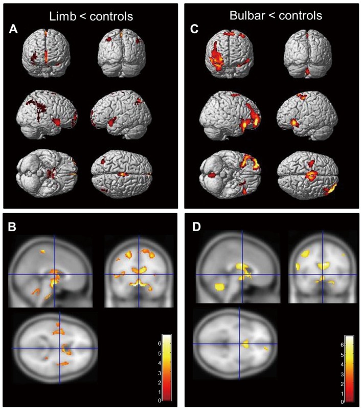
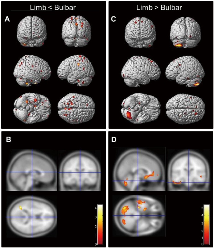
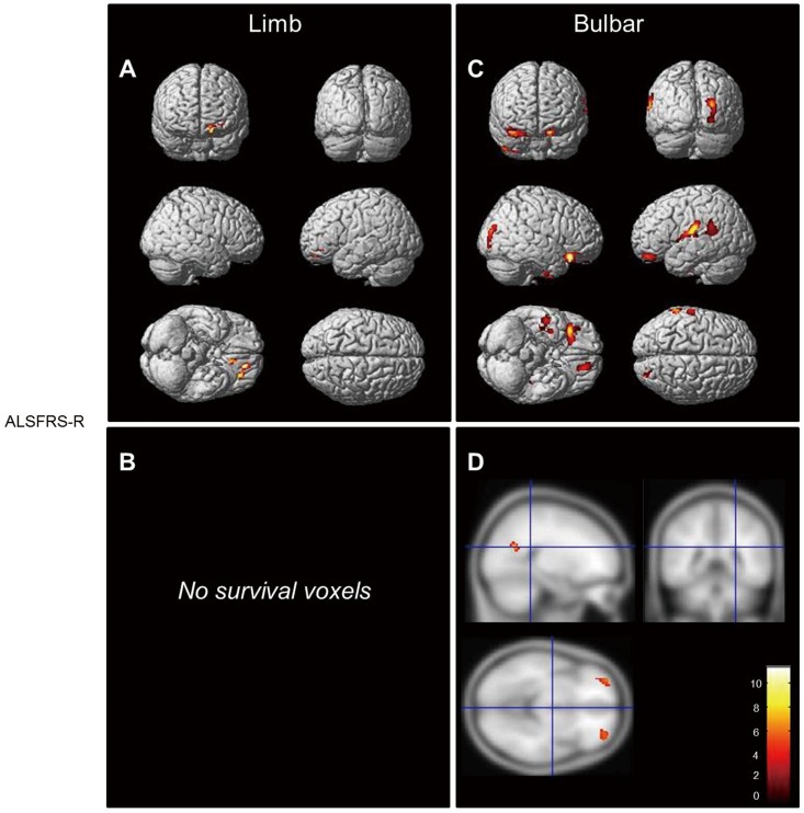
Results: In limb-onset ALS patients, atrophy was largely confined to the motor cortex and adjacent pre- and postcentral regions. However, in the bulbar-onset group, affected regions were more widespread and included these same areas but also extended to the bilateral frontotemporal and left superior temporal and supramarginal gyri, and multiple regression analysis revealed that their ALSFRS-R scores were associated with extensive loss of gray matter while FVC was related to atrophy in subcortical regions of the left superior temporal gyrus. In limb-onset ALS patients, disease duration was related to the degree of atrophy in the motor and adjacent areas.
Conclusion: Sporadic ALS subtypes show different patterns of brain atrophy. Neural networks related to limb and bulbar motor functions in each ALS subtype may underlie their distinct patterns of cerebral atrophy. That is, more extensive cortical and subcortical atrophy is correlated with greater ALSFRS-R severity and shorter disease duration in the bulbar-onset subtype and may explain the poor prognosis of these patients.
Conflict of interest statement
The authors have declared that no competing interests exist.
Figures
References
-
- Zufiria M, Gil-Bea FJ, Fernandez-Torron R, Poza JJ, Munoz-Blanco JL, Rojas-Garcia R, et al. ALS: A bucket of genes, environment, metabolism and unknown ingredients. Progress in neurobiology. 2016. Epub 2016/05/29. - PubMed
-
- Chio A, Logroscino G, Hardiman O, Swingler R, Mitchell D, Beghi E, et al. Prognostic factors in ALS: A critical review. Amyotrophic lateral sclerosis: official publication of the World Federation of Neurology Research Group on Motor Neuron Diseases. 2009;10(5–6):310–23. Epub 2009/11/20. - PMC - PubMed
Publication types
MeSH terms
Grants and funding
LinkOut - more resources
Full Text Sources
Other Literature Sources
Medical
Miscellaneous
