. 2017 Jan 17;13(1):e1006166.
doi: 10.1371/journal.ppat.1006166. eCollection 2017 Jan.

Replication-Coupled Recruitment of Viral and Cellular Factors to Herpes Simplex Virus Type 1 Replication Forks for the Maintenance and Expression of Viral Genomes

Affiliations

Replication-Coupled Recruitment of Viral and Cellular Factors to Herpes Simplex Virus Type 1 Replication Forks for the Maintenance and Expression of Viral Genomes

Jill A Dembowski et al. PLoS Pathog. .

Abstract

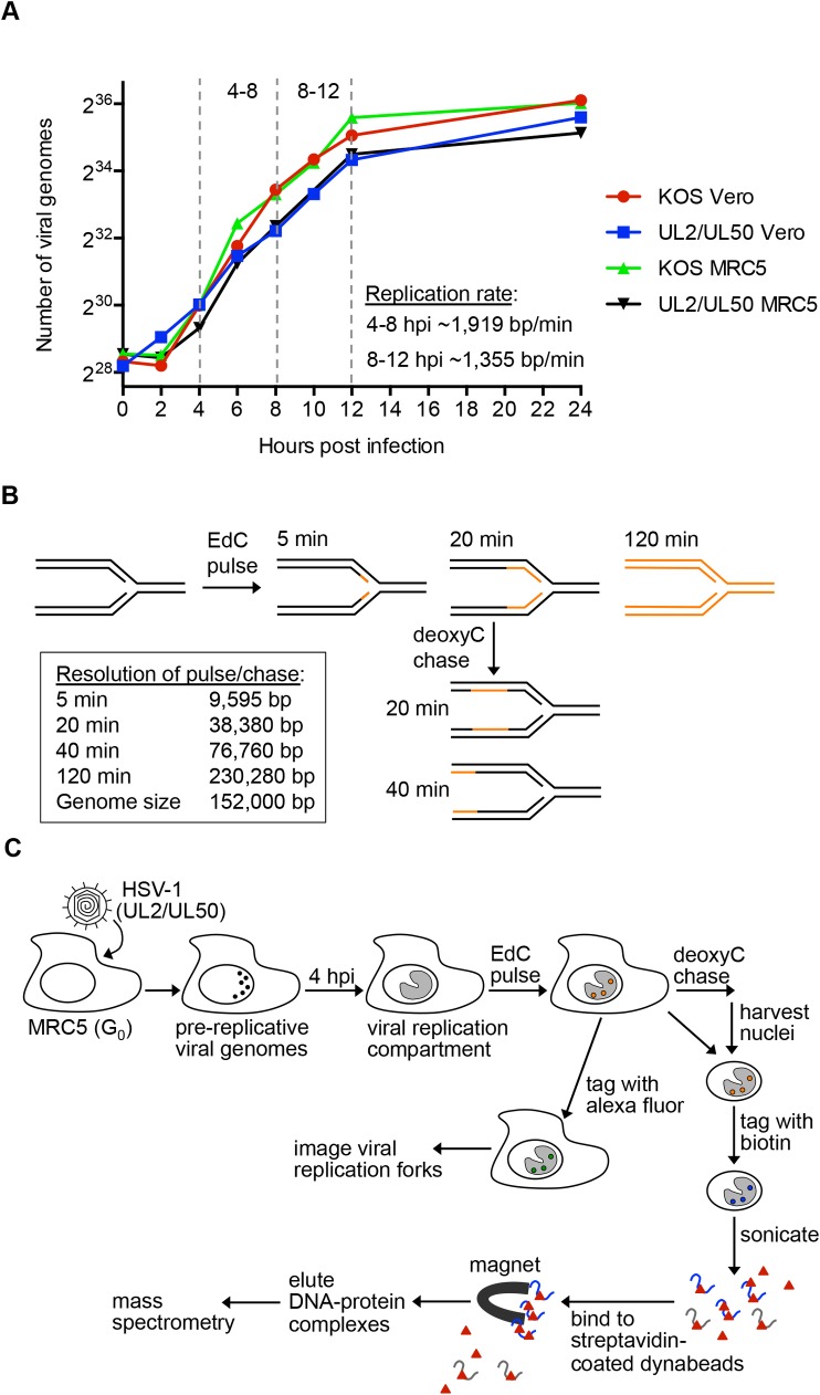
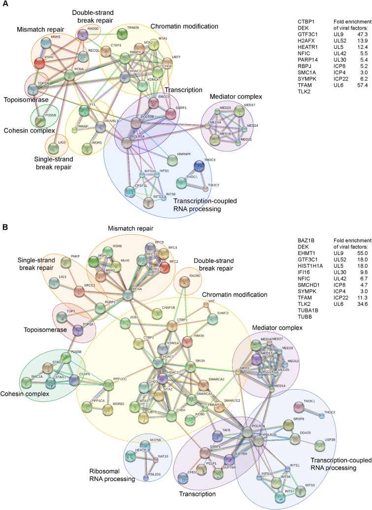
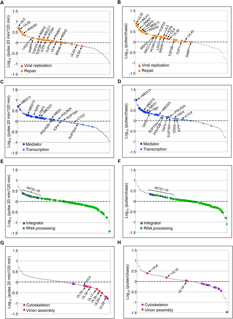
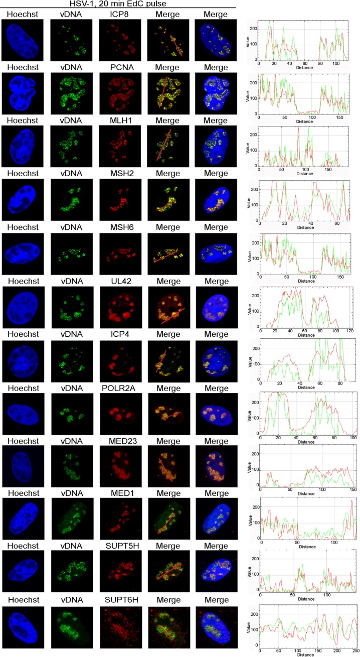
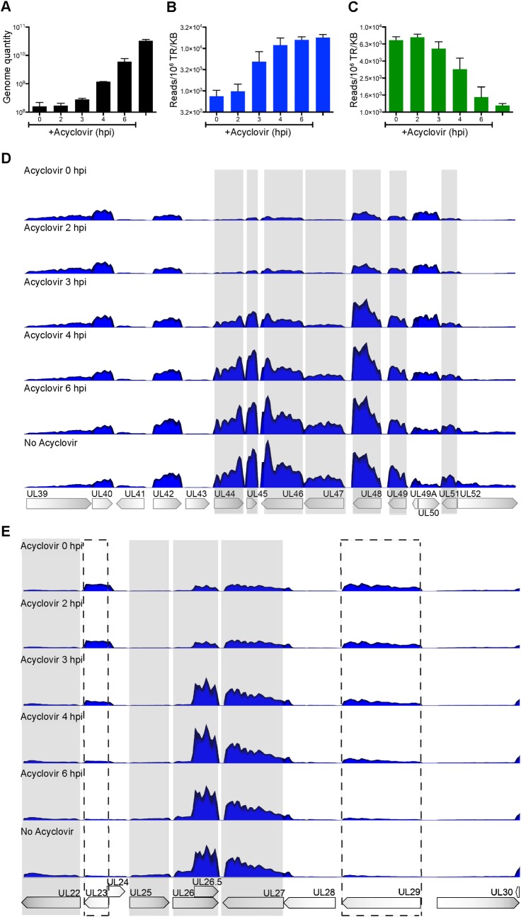
Herpes simplex virus type 1 (HSV-1) infects over half the human population. Much of the infectious cycle occurs in the nucleus of cells where the virus has evolved mechanisms to manipulate host processes for the production of virus. The genome of HSV-1 is coordinately expressed, maintained, and replicated such that progeny virions are produced within 4-6 hours post infection. In this study, we selectively purify HSV-1 replication forks and associated proteins from virus-infected cells and identify select viral and cellular replication, repair, and transcription factors that associate with viral replication forks. Pulse chase analyses and imaging studies reveal temporal and spatial dynamics between viral replication forks and associated proteins and demonstrate that several DNA repair complexes and key transcription factors are recruited to or near replication forks. Consistent with these observations we show that the initiation of viral DNA replication is sufficient to license late gene transcription. These data provide insight into mechanisms that couple HSV-1 DNA replication with transcription and repair for the coordinated expression and maintenance of the viral genome.


Conflict of interest statement

The authors have declared that no competing interests exist.

Figures

Fig 1
Fig 1. Methodology to Label, Image, and Purify HSV-1 Replication Forks.
A. Calculation of the rate of viral DNA replication. MRC5 or Vero cells were infected with KOS or UL2/UL50 and the number of viral genomes as a function of hours post infection (hpi) was determined by quantitative PCR (qPCR). The shown replication rates at 4–8 and 8–12 hpi were calculated using UL2/UL50 infected MRC5 cells because these conditions were used for subsequent proteomic analyses. B. Calculated resolution of pulse/chase experiments. Number of base pairs labeled or chased for various time intervals are indicated. C. Schematic of methods used to label and tag viral replication forks. Pre-replicative genomes coalesce and form replication compartments by 4 hpi. After pulse chase of viral replication forks, DNA was either tagged with an alexa fluor for imaging or biotin for purification following isolation of nuclei. For isolation of fork associated proteins, nuclei were lysed and DNA was fragmented by sonication and DNA-protein complexes were purified on streptavidin-coated beads. DNA protein complexes were eluted from beads after several wash steps (not depicted) and proteins were identified by mass spectrometry.
Fig 2
Fig 2. Viral Replication Proteins and Select Cellular Factors are Associated with HSV-1 Replication Forks.
A. STRING mapping of proteins enriched on viral replication forks after a 5 min EdC pulse. Human proteins enriched by 5 fold compared to the unlabeled negative control are shown in the functional interaction map or list of unmapped proteins, which was generated using STRING [28] with data settings to display only high confidence interactions. Gene names were used to map interactions. Circles indicate proteins that function in the same biological process. Fold enrichment of viral proteins is shown at right. B. STRING mapping of proteins enriched by 8 fold on viral replication forks after a 20 min pulse. See also S1 Table.
Fig 3
Fig 3. Pulse and Pulse Chase Analysis of Viral Replication Forks Reveals Interaction Dynamics Between Nascent Viral DNA and Associated Proteins.
Relative fork associated protein levels were compared between pulse labeled replication forks (20 min) and replicated genomes (120 min) (A, C, E, G) and between pulse labeled (pulse) and chased (chase) replication forks (B, D, F, H). Viral replication and cellular repair proteins (A, B), transcription factors (C, D), RNA processing factors (E, F), and cytoskeletal and virion assembly proteins (G, H) are highlighted in individual graphs. Gene names are indicated for corresponding proteins. See also S2 Fig and S1 Table.
Fig 4
Fig 4. Proteins Enriched on Viral Replication Forks Colocalize with Sites of Active Viral DNA Synthesis within HSV-1 Replication Compartments.
Sites of active viral DNA synthesis were labeled with EdC for 20 min after a four-hour infection of Vero cells with wild type HSV-1. Infected cells were fixed and EdC labeled DNA was tagged with alexa fluor 488 to visualize viral replication forks (green) and viral and cellular proteins were visualized by immunofluorescence (red). Nuclei were labeled with Hoechst (blue). Traces were generated using the RGB profiler plugin in Image J and correspond to the red line drawn on the red/green merge panel. Gene names that correspond to detected proteins are indicated for consistency with proteomics data. See also Table 1.
Fig 5
Fig 5. Initial Rounds of Replication are Sufficient for Late Gene Expression.
Vero cells were infected with wild type HSV-1 at an MOI of 10 PFU/cell and acyclovir was added to the growth medium as indicated at 0, 2, 3, 4, or 6 hpi. Total DNA or RNA was collected at 12 hpi. A. The number of viral genomes was measured by quantification of the gC gene by qPCR. Error bars represent the standard deviation of biological duplicates. B-E. The quantities of viral transcripts were measured using RNA-Seq. B. RNA-Seq analysis of the late gene UL44 (gC). The units on the y-axis are reads mapped to the viral gene locus per million total reads per kilobase pair (Reads/106 TR/KB). C. RNA-Seq analysis of the early gene UL23 (tk). D. Mapped reads are shown relative to the HSV-1 gene loci from UL39 to UL52. Each sample was normalized relative to 107 total reads. The y-axis maximum value is 75,000 reads. The gray shaded areas indicate late genes. E. Mapped reads are shown relative to the KOS gene loci map from UL22 to UL30. Dashed boxes indicate early genes UL23 (tk) and UL29 (ICP8).
Fig 6
Fig 6. Model Illustrating HSV-1 Replication-Coupled Processes.
A. Subdomains within HSV-1 replication compartments contain genomes participating in replication-coupled and replication-independent processes. B. Model depicting replication-coupled DNA repair and C. transcription.

References

    1. Smith S, Weller SK (2015) HSV-I and the cellular DNA damage response. Future Virol 10: 383–397. 10.2217/fvl.15.18 - DOI - PMC - PubMed
    1. Roizman B, Zhou G (2015) The 3 facets of regulation of herpes simplex virus gene expression: A critical inquiry. Virology 479–480: 562–567. 10.1016/j.virol.2015.02.036 - DOI - PMC - PubMed
    1. McGeoch DJ, Dalrymple MA, Davison AJ, Dolan A, Frame MC, et al. (1988) The complete DNA sequence of the long unique region in the genome of herpes simplex virus type 1. J Gen Virol 69 (Pt 7): 1531–1574. - PubMed
    1. McGeoch DJ, Dolan A, Donald S, Rixon FJ (1985) Sequence determination and genetic content of the short unique region in the genome of herpes simplex virus type 1. J Mol Biol 181: 1–13. - PubMed
    1. Alwine JC, Steinhart WL, Hill CW (1974) Transcription of herpes simplex type 1 DNA in nuclei isolated from infected HEp-2 and KB cells. Virology 60: 302–307. - PubMed

MeSH terms