. 2017 Jan 17;11(1):e0005285.
doi: 10.1371/journal.pntd.0005285. eCollection 2017 Jan.

Cytoplasmic Localization of HTLV-1 HBZ Protein: A Biomarker of HTLV-1-Associated Myelopathy/Tropical Spastic Paraparesis (HAM/TSP)

Affiliations

Cytoplasmic Localization of HTLV-1 HBZ Protein: A Biomarker of HTLV-1-Associated Myelopathy/Tropical Spastic Paraparesis (HAM/TSP)

Marco Baratella et al. PLoS Negl Trop Dis. .

Abstract

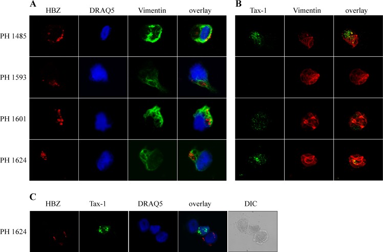
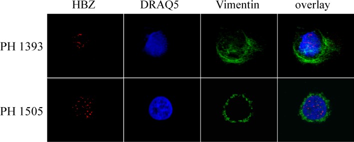
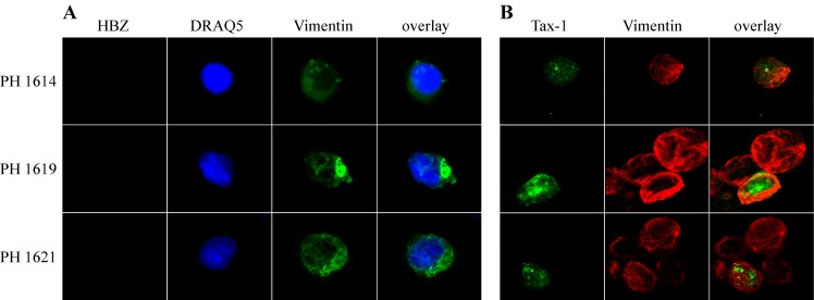
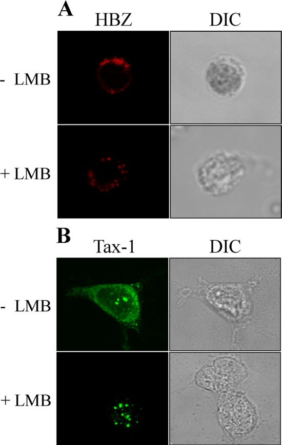
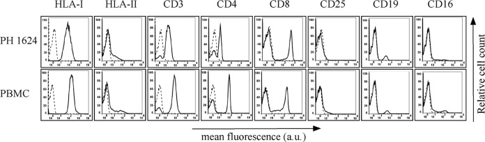
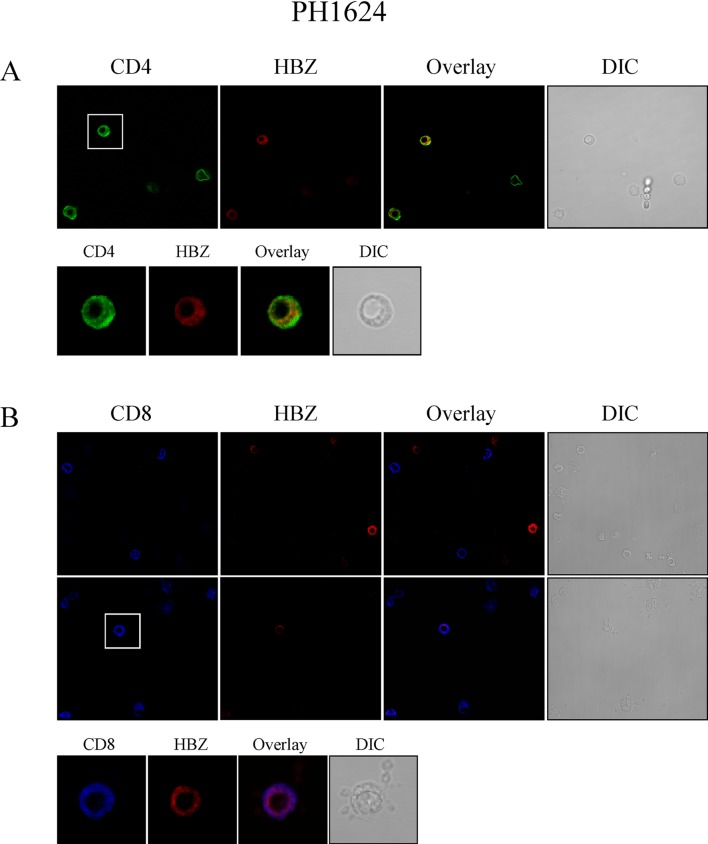
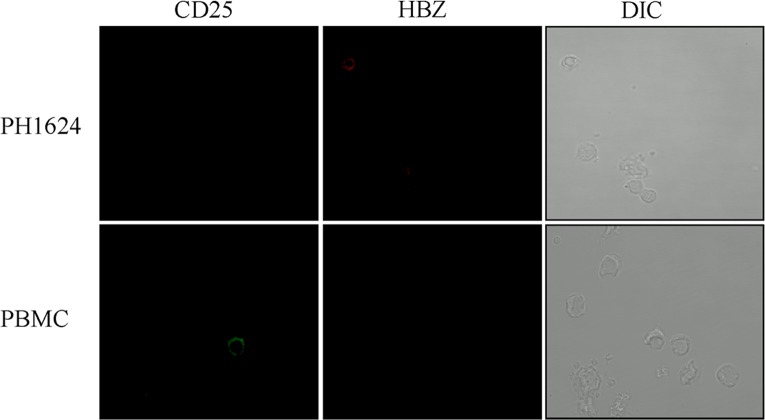
HTLV-1 is the causative agent of a severe form of adult T cell leukemia/Lymphoma (ATL), and of a chronic progressive neuromyelopathy designated HTLV-1 associated myelopathy/tropical spastic paraparesis (HAM/TSP). Two important HTLV-1-encoded proteins, Tax-1 and HBZ, play crucial roles in the generation and maintenance of the oncogenic process. Less information is instead available on the molecular and cellular mechanisms leading to HAM/TSP. More importantly, no single specific biomarker has been described that unambiguously define the status of HAM/TSP. Here we report for the first time the finding that HBZ, described until now as an exclusive nuclear protein both in chronically infected and in ATL cells, is instead exclusively localized in the cytoplasm of peripheral blood mononuclear cells (PBMC) from patients suffering of HAM/TSP. Interestingly, at the single cell level, HBZ and Tax-1 proteins are never found co-expressed in the same cell, suggesting the existence of mechanisms of expression uncoupling of these two important HTLV-1 viral products in HAM/TSP patients. Cells expressing cytoplasmic HBZ were almost exclusively found in the CD4+ T cell compartment that was not, at least in a representative HAM/TSP patient, expressing the CD25 marker. Less than 1 percent CD8+ T cells were fond positive for HBZ, while B cells and NK cells were found negative for HBZ in HAM/TSP patients. Our results identify the cytoplasmic localization of HBZ in HAM/TSP patient as a possible biomarker of this rather neglected tropical disease, and raise important hypotheses on the role of HBZ in the pathogenesis of the neuromyelopathy associated to HTLV-1 infection.


Conflict of interest statement

The authors have declared that no competing interests exist

Figures

Fig 1
Fig 1. Subcellular localization of endogenous HBZ and Tax-1 in PBMC of HAM/TSP patients.
(A) PBMC of four HAM/TSP patients (PH1485, PH1593, PH1601, and PH1624) were stained with the 4D4-F3 anti-HBZ mAb followed by Alexa Fluor 546-conjugated goat anti-mouse IgG1 antibody (red) and (B) with the A51-2 anti-Tax-1 mAb followed by Alexa Fluor 488-conjugated goat-anti-mouse IgG2a antibody (green), and analyzed by confocal microscopy. Specific counterstaining of nucleus or cytoplasmic compartments was performed by using DRAQ5 fluorescence probe to detect the nucleus and anti-vimentin rabbit polyclonal antibody followed by goat anti-rabbit IgG conjugated to Alexa Fluor 488 (green, panel A) or to Alexa Fluor 546 (red, panel B) to stain the cytoplasmic compartment. At least 300 cells were analyzed; a representative cell for HBZ or for Tax-1 staining is shown for each patient.(C) Low magnification field to show the co-existence of cells mutually exclusive for the expression of cytoplasmic HBZ and Tax-1 in PBMC of patient PH1624. Cells were co-stained with 4D4-F3 anti-HBZ mAb and with A51-2 anti-Tax-1 mAb followed by specific secondary antibodies staining as described in (A) and (B).
Fig 2
Fig 2. Subcellular localization of endogenous HBZ and Tax-1 in PBMC of ATL patients.
PBMC of two ATL patients (PH1393 and PH1505) were stained with the anti-HBZ 4D4-F3 mAb, followed by Alexa Fluor 546-conjugated goat anti-mouse IgG1antibody to detect the HBZ protein and analyzed by confocal microscopy. Specific counterstaining of nucleus or cytoplasmic compartments was performed by using DRAQ5 fluorescence probe or anti-vimentin antibody as describe in the legend to Fig 1. At least 300 cells were analyzed; a representative cell for HBZ staining is shown for each patient. PBMC from both patients were negative for expression of Tax-1 protein.
Fig 3
Fig 3. Lack of endogenous HBZ, but not of Tax-1, detection in PBMC of HTLV-1 positive asymptomatic carriers.
(A) PBMC of three HTLV-1-positive asymptomatic carriers (PH1614, PH1619, and PH1621) were stained with the anti-HBZ 4D4-F3 and (B) the anti-Tax-1 A51-2 mAbs, followed by Alexa Fluor 546-conjugated goat anti-mouse IgG1antibody to detect HBZ protein (red), or by Alexa Fluor 488-conjugated goat-anti-mouse IgG2a to detect Tax-1 (green), and analyzed by confocal microscopy. Specific counterstaining of nucleus or cytoplasmic compartments was performed by using DRAQ5 fluorescence probe to detect the nucleus and anti-vimentin rabbit polyclonal antibody followed by goat anti-rabbit IgG conjugated to Alexa Fluor 488 (green, panel A) or to Alexa Fluor 546 (red, panel B) to stain the cytoplasmic compartment. At least 300 cells were analyzed; a representative cell is shown for each patient.
Fig 4
Fig 4. HBZ is a resident cytoplasmic protein and does not shuttle to the nucleus in HAM/TSP.
(A) PBMC of HAM/TSP patient PH1624 were either treated (+ LMB) or untreated (- LMB) with LMB, an inhibitor of nuclear export, before fixing and staining with the anti-HBZ 4D4-F3 mAb followed by Alexa Fluor 546-conjugated goat anti-mouse IgG1antibody (red). (B) As control of inhibition of nuclear export by LMB, 293T cells transfected with the plasmid coding for Tax-1 viral protein were treated with LMB (+LMB) or left untreated (-LMB). Cells were fixed and stained with A51-2 anti Tax-1 mAb followed by Alexa Fluor 488-conjugated goat-anti-mouse IgG2a (green), and analyzed by confocal microscopy. DIC represents the differential interference contrast image.
Fig 5
Fig 5. Expression of cells surface markers in PBMC of HAM/TSP patient PH1624 and healthy control.
The expression of HLA class I, HLA class II DR, CD3, CD4, CD8, CD25, CD19, and CD16 surface molecules on PBMC from TSP/HAM patient PH1624 and a healthy control was assessed by immunofluorescence and flow cytometry with antibodies specific for the various markers. Results are expressed as relative number of cells (ordinate) versus the mean intensity of fluorescence in arbitrary units (abscissa). In each histogram, negative controls, obtained by staining the cells with an appropriate isotype-matched antibody, are depicted as dashed line.
Fig 6
Fig 6. HBZ is preferentially expressed in CD4+ T cells of HAM/TSP patient PH1624.
Confocal microscopy analysis of PBMC from HAM/TSP patient PH1624. (A) co-staining with the 4D4-F3 anti-HBZ mAb followed by Alexa Fluor 546-conjugated goat anti-mouse IgG1 antibody (red) and with the anti-CD4 mAb followed by Alexa Fluor 488-conjugated goat-anti-rabbit IgG antibody (green); upper panels, extended field; lower panels, enlarged field focused on the single cell depicted in the square of the left upper panel and positive for both CD4 and HBZ. (B) co-staining with the 4D4-F3 anti-HBZ mAb followed by Alexa Fluor 546-conjugated goat anti-mouse IgG1 antibody (red) and with the anti-CD8 rabbit monoclonal antibody directly conjugated to Alexa Fluor 647 (blue); upper panels depict extended fields with CD8+ cells negative for HBZ; middle panels, extended fields with many CD8+ cells negative for HBZ and the single CD8+/HBZ+ cell (square); lower panels, enlarged field on the single CD8+/HBZ+ cell.
Fig 7
Fig 7. Lack of CD25+ cell in PBMC of patient PH1624, as assessed by confocal microscopy.
PBMC of HAM/TSP patient PH1624 and of healthy control were co-stained with the anti-HBZ 4D4-F3 mAb followed by Alexa Fluor 546-conjugated goat anti-mouse IgG1 antibody (red) and with the anti-CD25 monoclonal antibody directly conjugated to Alexa Fluor 488 (green) and analyzed by confocal microscopy. Upper left panel shows the negativity of CD25 staining in PH1624 patient as compared with a positive cell for the same marker in PBMC of healthy control (PBMC, lower right panel)

Similar articles

Cited by

References

    1. Gessain A, Cassar O. Epidemiological aspects and world distribution of HTLV-1 infection. Frontiers Microbiol. 2012;3: 388. - PMC - PubMed
    1. Poiesz B, Ruscetti F, Gazdar A, Bunn P, Minna J, Gallo R. Detection and isolation of type C retrovirus particles from fresh and cultured lymphocytes of a patient with cutaneous T-cell lymphoma Proc Natl Acad Sci USA. 1980;77:7415–9. - PMC - PubMed
    1. Gessain A, Barin F, Vernant JC, Gout O, Maurs L, Calender A, et al. Antibodies to human T-lymphotropic virus type-I in patients with tropical spastic paraparesis. Lancet. 1985;2:407–10. - PubMed
    1. Osame M, Usuku K, Izumo S, Ijichi N, Amitani H, Igata A, et al. HTLV-I associated myelopathy, a new clinical entity. Lancet. 1986;1:1031–2. - PubMed
    1. Matsuoka M, Jeang KT. Human T-cell leukaemia virus type 1 (HTLV-1) infectivity and cellular transformation. Nat Rev Cancer. 2007;7:270–80. 10.1038/nrc2111 - DOI - PubMed

Publication types

MeSH terms

Substances