. 2017 Jan 17;12(1):e0170154.
doi: 10.1371/journal.pone.0170154. eCollection 2017.

Changes in Circulating B Cell Subsets Associated with Aging and Acute SIV Infection in Rhesus Macaques

Affiliations

Changes in Circulating B Cell Subsets Associated with Aging and Acute SIV Infection in Rhesus Macaques

W L William Chang et al. PLoS One. .

Abstract

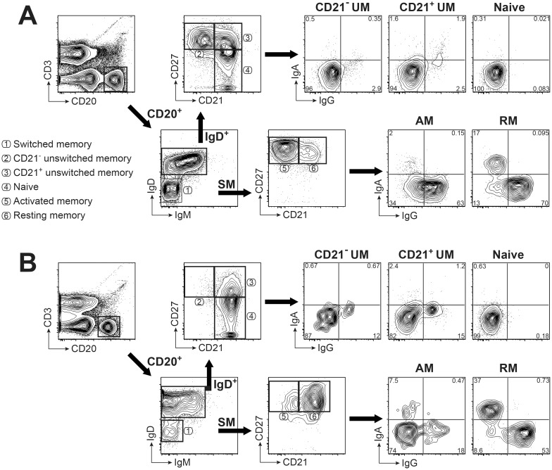
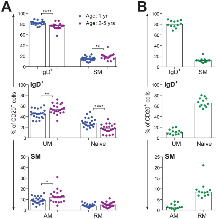
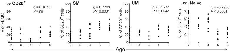
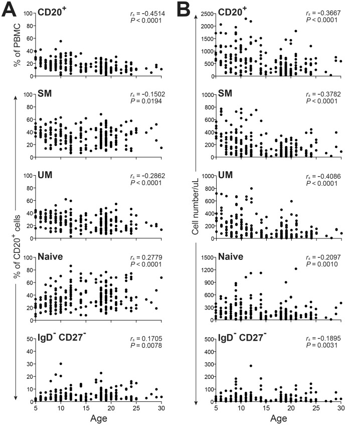
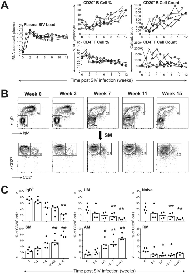
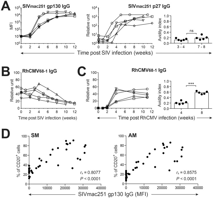
Aging and certain viral infections can negatively impact humoral responses in humans. To further develop the nonhuman primate (NHP) model for investigating B cell dynamics in human aging and infectious disease, a flow cytometric panel was developed to characterize circulating rhesus B cell subsets. Significant differences between human and macaque B cells included the proportions of cells within IgD+ and switched memory populations and a prominent CD21-CD27+ unswitched memory population detected only in macaques. We then utilized the expanded panel to analyze B cell alterations associated with aging and acute simian immunodeficiency virus (SIV) infection in the NHP model. In the aging study, distinct patterns of B cell subset frequencies were observed for macaques aged one to five years compared to those between ages 5 and 30 years. In the SIV infection study, B cell frequencies and absolute number were dramatically reduced following acute infection, but recovered within four weeks of infection. Thereafter, the frequencies of activated memory B cells progressively increased; these were significantly correlated with the magnitude of SIV-specific IgG responses, and coincided with impaired maturation of anti-SIV antibody avidity, as previously reported for HIV-1 infection. These observations further validate the NHP model for investigation of mechanisms responsible for B cells alterations associated with immunosenescence and infectious disease.


Conflict of interest statement

The authors have declared that no competing interests exist.

Figures

Fig 1
Fig 1. Gating strategy for identification of memory B cell subsets in rhesus macaque and human PBMC.
B cell phenotyping panels with the same markers and gating strategy are illustrated for macaque (A) and human (B) PBMC. Shown are 5% contour plots with outliers of cells after gating on singlets, then lymphocytes, and exclusion of dead cells. B cells were identified by gating on CD3- CD20+ cells. Naïve and unswitched memory (UM) B cells were defined using CD21 and CD27 after gating on IgD+ B cells. Activated memory (AM) and resting memory (RM) B cells were defined by using CD21 and CD27 in the IgD- IgM- switched memory (SM) B cell subset. Surface expression of IgG and IgA on each B cell subset is shown in the right panel. Numbers represent percentages of the respective cell populations among the total gated cells. Circled numbers associated with different gated populations identify the B cell subset within that gate as noted in the list of subsets.
Fig 2
Fig 2. Characterization of isotype expressing memory B cells by combining surface and intracellular staining for IgG and IgA.
(A) Representative FACS plots of circulating B cell subsets staining for IgG and IgA are shown. Numbers represent percentages of the respective cell populations among the total gated cells. Anti-IgG and/or Anti-IgA antibodies were added before (surface) or after (surface + intracellular) membrane permeablilization. (B) Comparison of IgG+ or IgA+ expression by AM or RM B cells by surface only (s) or surface plus intracellular (s + i) staining with isotype-specific antibodies. Statistical analyses were performed using nonparametric Wilcoxon tests. ns: not significant.
Fig 3
Fig 3. Distribution of B cell subsets in PBMC.
(A) Frequencies of B cell subsets in peripheral blood of healthy SPF rhesus macaques (age 1 yr, n = 23; age 2 − 5 yrs, n = 21) are shown, with each data point representing an individual animal. (B) Frequencies of B cell subsets in peripheral blood of healthy human adults (n = 12) are shown, with each data point representing an individual donor. Bars in each graph indicate median frequencies. Statistical analyses between two age cohorts of SPF macaques were performed using nonparametric Mann-Whitney tests. Symbol: * P < 0.05; ** P < 0.01; **** P < 0.0001.
Fig 4
Fig 4. Frequencies of total CD20+ B cells and B cell subsets in juvenile and young adult non-SPF rhesus macaques.
PBMC isolated from non-SPF macaques (age 2 − 6 yrs, n = 50) were assessed by FACS to determine the frequencies of CD20+ B cells, and SM, UM, and naïve, B cell subsets. Correlations between age and cell frequencies were determined by Spearman’s rank correlation.
Fig 5
Fig 5. Frequencies and absolute cell numbers of circulating B cells in young adult, adult, and aged non-SPF rhesus macaques.
PBMC isolated from non-SPF macaques distinguished as young adult and adult (age 5 − 18 yrs, n = 158) and aged (age >18 yrs, n = 84) were assessed by FACS to determine the frequencies (A) and absolute cell counts (B) of CD20+ B cells, and SM, UM, Naïve, IgD-CD27- B cell subsets over time according to age. Correlations between age and cell frequencies were determined by Spearman’s rank correlation.
Fig 6
Fig 6. Impact of SIV infection on distribution of peripheral blood B cell subsets in rhesus macaques.
(A) Measurement of viral loads, B and CD4+ T cell frequencies and counts during acute and early set point SIV infection are shown. (B) Representative FACS plots depict the progressive shift of circulating B cell subsets over the duration of SIV infection. (C) Frequencies of peripheral blood B cell subsets in typical progressors (n = 5) before and after SIV infection are shown with each data point representing an individual animal. Bars in each graph indicate median frequencies. Statistical analyses were performed using Friedman nonparametric tests followed by Dunn’s multiple comparison tests comparing each time point with the week 0 control. Symbol: * P < 0.05; ** P < 0.01.
Fig 7
Fig 7. Comparison of antiviral antibody kinetics following SIV and RhCMV infection.
(A) Plasma titers of anti-SIV gp130 and p27 IgG during SIV infection are shown. Changes in anti-SIV p27 avidity indices post SIV infection are shown on the right. (B) SIV infection impairs RhCMV-specific humoral immune responses. (C) Changes in plasma anti-RhCMV IgG titers and avidity indices are noted during primary RhCMV infection. (D) Correlation of SM and AM B cell frequencies in PBMC of SIV-infected macaques with plasma antibody titers of anti-SIV gp130 was determined. Statistical analyses of avidities indices were performed using Student’s t tests (paired, two-tailed). Correlation analyses were performed by Spearman’s rank tests. Symbol: *** P < 0.001. ns: not significant.

References

    1. Pieper K, Grimbacher B, Eibel H. B-cell biology and development. J Allergy Clin Immunol. 2013;131: 959–971. 10.1016/j.jaci.2013.01.046 - DOI - PubMed
    1. Chidrawar S, Khan N, Wei W, McLarnon A, Smith N, Nayak L, et al. Cytomegalovirus-seropositivity has a profound influence on the magnitude of major lymphoid subsets within healthy individuals. Clin Exp Immunol. 2009;155: 423–432. 10.1111/j.1365-2249.2008.03785.x - DOI - PMC - PubMed
    1. Goldeck D, Oettinger L, Janssen N, Demuth I, Steinhagen-Thiessen E, Pawelec G. Cytomegalovirus Infection Minimally Affects the Frequencies of B-Cell Phenotypes in Peripheral Blood of Younger and Older Adults. Gerontology. 2016;62: 323–329. 10.1159/000382076 - DOI - PubMed
    1. Wang C, Liu Y, Xu LT, Jackson KJ, Roskin KM, Pham TD, et al. Effects of aging, cytomegalovirus infection, and EBV infection on human B cell repertoires. J Immunol. 2014;192: 603–611. 10.4049/jimmunol.1301384 - DOI - PMC - PubMed
    1. Amu S, Ruffin N, Rethi B, Chiodi F. Impairment of B-cell functions during HIV-1 infection. AIDS. 2013;27: 2323–2334. 10.1097/QAD.0b013e328361a427 - DOI - PubMed

MeSH terms