

Xenopus as a model for studies in mechanical stress and cell division
- PMID: 28095623
- PMCID: PMC5276720
- DOI: 10.1002/dvg.23004
Xenopus as a model for studies in mechanical stress and cell division
Abstract
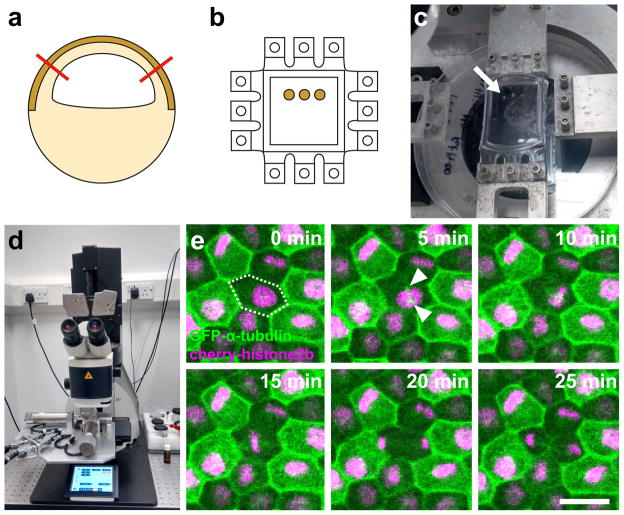
We exist in a physical world, and cells within biological tissues must respond appropriately to both environmental forces and forces generated within the tissue to ensure normal development and homeostasis. Cell division is required for normal tissue growth and maintenance, but both the direction and rate of cell division must be tightly controlled to avoid diseases of over-proliferation such as cancer. Recent studies have shown that mechanical cues can cause mitotic entry and orient the mitotic spindle, suggesting that physical force could play a role in patterning tissue growth. However, to fully understand how mechanics guides cells in vivo, it is necessary to assess the interaction of mechanical strain and cell division in a whole tissue context. In this mini-review we first summarise the body of work linking mechanics and cell division, before looking at the advantages that the Xenopus embryo can offer as a model organism for understanding: (1) the mechanical environment during embryogenesis, and (2) factors important for cell division. Finally, we introduce a novel method for applying a reproducible strain to Xenopus embryonic tissue and assessing subsequent cell divisions.
Keywords: Xenopus laevis; biomechanics; division orientation; mitosis.
© 2017 Wiley Periodicals, Inc.
Figures
References
-
- Baena-Lopez LA, Baonza A, Garcia-Bellido A. The orientation of cell divisions determines the shape of Drosophila organs. Curr Biol. 2005;15:1640–1644. - PubMed
-
- Beloussov LV, Dorfman JG, Cherdantzev VG. Mechanical stresses and morphological patterns in amphibian embryos. J Embryol Exp Morphol. 1975;34:559–574. - PubMed
Publication types
MeSH terms
Grants and funding
LinkOut - more resources
Full Text Sources
Other Literature Sources
