Review
. 2017 Jan;55(1-2):10.1002/dvg.23002.
doi: 10.1002/dvg.23002.

A frog's view of EphrinB signaling

Affiliations
Review

A frog's view of EphrinB signaling

Yoo-Seok Hwang et al. Genesis. 2017 Jan.

Abstract

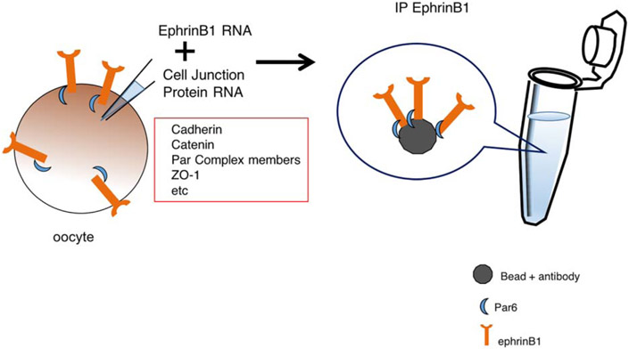
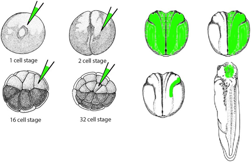
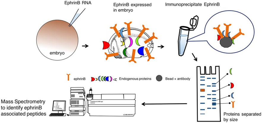
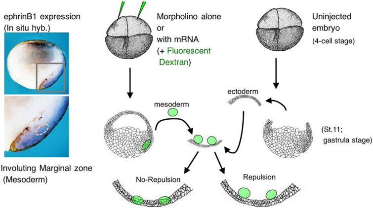
Cell-cell and cell-substrate adhesion are essential to the proper formation and maintenance of tissue patterns during development, and deregulation of these processes can lead to invasion and metastasis of cancer cells. Cell surface adhesion and signaling molecules are key players in both normal development and cancer progression. One set of cell surface proteins, the Eph receptor tyrosine kinases and their membrane-bound ligands, ephrins, are significant regulators of these processes. During embryonic development, the Eph/ephrin signaling system is involved in cell-cell contact events that result in cell sorting and boundary formation between receptor and ligand bearing cells. When migrating cells that display the membrane bound ligands or receptors come in contact with cells bearing the cognate partner, the response may be adhesion or repulsion, ultimately leading to the proper positioning of these cells. During cancer progression, the signaling between these receptor/ligand pairs is often deregulated, leading to increased invasion and metastasis. To gain mechanistic insight into the pathways that mediate Eph receptor and ephrin signaling we have relied upon a very tractable system, the frog Xenopus. This model system has proven to be extremely versatile, and represents a relatively quick and manipulable system to explore signaling events and the in vivo processes affected by these signals.

Keywords: Eph; Xenopus; development.


Figures

FIGURE 1
FIGURE 1
A limited screen for candidate interacting proteins. This schematic depicts the use of isolated stage 6 Xenopus oocytes to test whether a protein of interest (ephrinB1) is able to interact with possible candidate proteins (cell–cell adhesion and cell boundary proteins). Oocytes are injected with in vitro transcribed mRNA encoding ephrinB1 and individual candidate gene mRNAs, cultured for several hours, lysates prepared and ephrinB1 immune-complexes collected. Western analysis should then be performed to detect whether a candidate associates with the ephrinB1 protein
FIGURE 2
FIGURE 2
Schematic depicting spatial control of mRNA or MO location in a Xenopus embryo. GFP mRNA or fluorescent dye can be injected along with a particular mRNA or MO in specific blastomeres at various stages of development to spatially restrict protein expression or knockdown, respectively. The target area can be assessed by epifluorescence microscopy
FIGURE 3
FIGURE 3
Schematic depicting the use of immunoprecipitation and mass spectrometry to identify possible ephrinB-associated proteins. Xenopus embryos are injected with ephrinB mRNA, cultured until later stages, lysates prepared, and ephrinB immune-complexes collected. Proteins are separated by gel electrophoresis, gel slices removed and prepared for mass spectrometric analysis to identify proteins in the immune-complex
FIGURE 4
FIGURE 4
Use of explants to determine the role of proteins in tissue repulsion. The left side of the figure shows a wholemount in situ hybridization of a bisected Xenopus embryo with a probe for ephrinB1 expression; showing expression in the involuting marginal zone. The right side is a schematic depicting the BCR/mesoderm repulsion assay. Four cell stage embryos are injected with either mRNA or MOs or both, along with fluorescent dextran as a tracer. The mesoderm (involuting marginal zone) is removed and placed upon the blastocoel roof (BCR) explant from an uninjected embryo. Light and epifluorescence microscopy allows for visualization of whether the two explants mix or repulse one another

References

    1. Adams RH, Diella F, Hennig S, Helmbacher F, Deutsch U, & Klein R (2001). The cytoplasmic domain of the ligand ephrinB2 is required for vascular morphogenesis but not cranial neural crest migration. Cell, 104, 57–69. - PubMed
    1. Banach M, Edholm ES, & Robert J (2016). Exploring the functions of nonclassical MHC class Ib genes in Xenopus laevis by the CRISPR/Cas9 system. Developmental Biology. doi: 10.1016/j.ydbio.2016.05.023. [Epub ahead of print] - DOI - PMC - PubMed
    1. Bhattacharya D, Marfo CA, Li D, Lane M, & Khokha MK (2015) CRISPR/Cas9: An inexpensive, efficient loss of function tool to screen human disease genes in Xenopus. Developmental Biology 408,196 – 204. - PMC - PubMed
    1. Blitz IL, Biesinger J, Xie X, & Cho KW (2013). Biallelic genome modification in F(0) Xenopus tropicalis embryos using the CRISPR/Cas system. Genesis, 51, 827–834. - PMC - PubMed
    1. Blitz IL, Fish MB, & Cho KW (2016). Leapfrogging: primordial germ cell transplantation permits recovery of CRISPR/Cas9-induced mutations in essential genes. Development, 143, 2868–2875. - PMC - PubMed

Publication types

LinkOut - more resources