

Relationship of diagnostic accuracy of renal cortical echogenicity with renal histopathology in dogs and cats, a quantitative study
- PMID: 28095845
- PMCID: PMC5240265
- DOI: 10.1186/s12917-016-0941-z
Relationship of diagnostic accuracy of renal cortical echogenicity with renal histopathology in dogs and cats, a quantitative study
Abstract
Background: Renal cortical echogenicity is routinely evaluated during ultrasonographic investigation of the kidneys. Both in dog and cat previous ex-vivo studies have revealed a poor correlation between renal echogenicity and corresponding lesions. The aim of this study was to establish the in-vivo relationship between renal cortical echogenicity and renal histopathology.
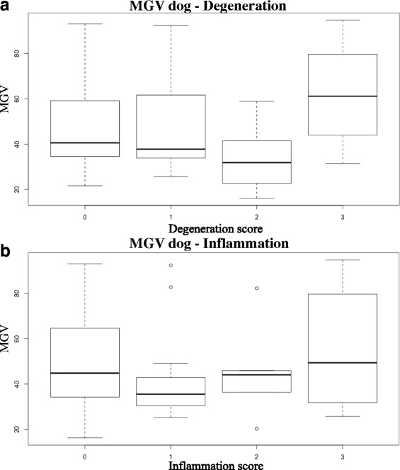
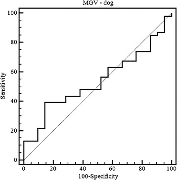
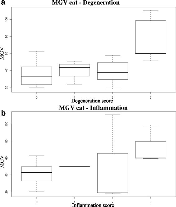
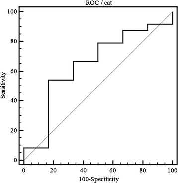
Results: Thirty-eight dogs and fifteen cats euthanized for critical medical conditions were included in the study. Ultrasonographic images of both kidneys were acquired ante mortem at standardized ultrasonographic settings. The echogenicity was quantified by means of Mean Gray Value (MGV) of the renal cortex measured with ImageJ. A complete histopathological examination of both kidneys was performed. Five kidneys were excluded because histopathology revealed neoplastic lesions. Only samples affected by tubular atrophy showed statistically different values in dog, and histopathology explained 13% of the total variance. MGV was not correlated neither to the degeneration nor to the inflammation scores. However, significant differences were identified between mildly and severely degenerated samples. Overall, the classification efficiency of MGV to detect renal lesions was poor with a sensitivity of 39% and a specificity of 86%. In cats, samples affected by both tubular vacuolar degeneration and interstitial nephritis were statistically different and histopathology explained 44% of the total variance. A linear correlation was evident between degeneration and MGV, whereas no correlation with inflammation was found. Statistically significant differences were evident only between normal and severely degenerated samples with a sensitivity of 54.17% and a specificity of 83.3% and MGV resulted scarce to discriminate renal lesions in this species.
Conclusions: Renal cortical echogenicity shows low relevance in detecting chronic renal disease in dog whereas it results worth to identify severe renal damage in cat.
Keywords: Image analysis; Kidney; Pathology; Ultrasound.
Figures
References
-
- D’Anjou MA, Penninck D. Kidneys and ureters. In: D’Anjou MA, Penninck D, editors. Atlas Small Animal Ultrasound, Second Ed. Ames: Wiley Blackwell; 2015. pp. 331–361.
-
- Nyland T, Widmer W, Mattoon JS. Urinary Tract. In: Nyland T, Mattoon JS, editors. Small Animal Diagnostic Ultrasound. 3. St. Luis: Elsevier; 2015. pp. 557–607.
MeSH terms
LinkOut - more resources
Full Text Sources
Other Literature Sources
Medical
Miscellaneous
