. 2017 Mar 23;129(12):1669-1679.
doi: 10.1182/blood-2016-08-733519. Epub 2017 Jan 17.

Platelets activate a pathogenic response to blood-stage Plasmodium infection but not a protective immune response

Affiliations

Platelets activate a pathogenic response to blood-stage Plasmodium infection but not a protective immune response

Irene Gramaglia et al. Blood. .

Abstract

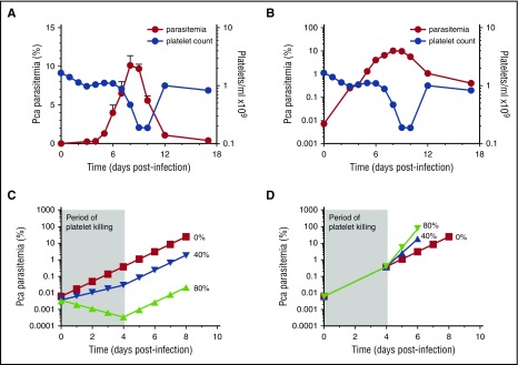
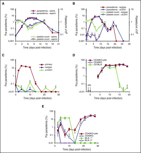
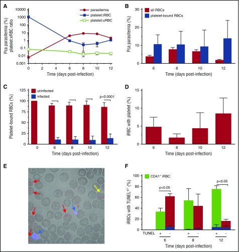
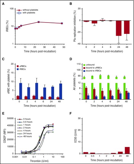
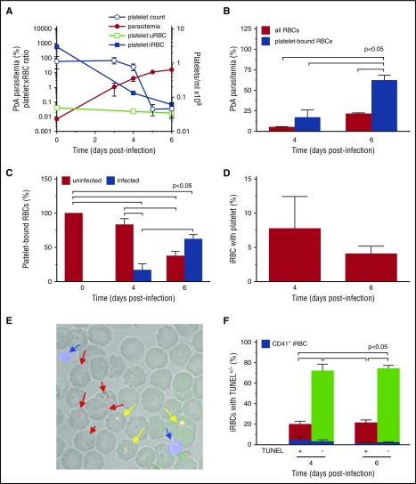
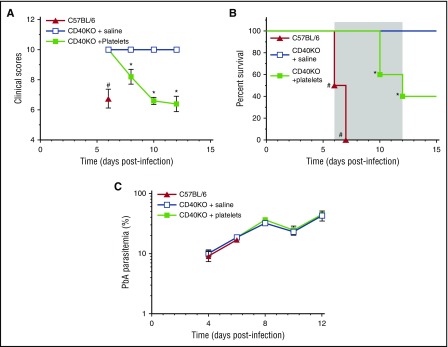
Clinical studies indicate that thrombocytopenia correlates with the development of severe falciparum malaria, suggesting that platelets either contribute to control of parasite replication, possibly as innate parasite killer cells or function in eliciting pathogenesis. Removal of platelets by anti-CD41 mAb treatment, platelet inhibition by aspirin, and adoptive transfer of wild-type (WT) platelets to CD40-KO mice, which do not control parasite replication, resulted in similar parasitemia compared with control mice. Human platelets at a physiologic ratio of 1 platelet to 9 red blood cells (RBCs) did not inhibit the in vitro development or replication of blood-stage Plasmodium falciparum The percentage of Plasmodium-infected (iRBCs) with bound platelets during the ascending parasitemia in Plasmodium chabaudi- and Plasmodium berghei-infected mice and the 48-hour in vitro cycle of P falciparum was <10%. P chabaudi and P berghei iRBCs with apoptotic parasites (TdT+) exhibited minimal platelet binding (<5%), which was similar to nonapoptotic iRBCs. These findings collectively indicate platelets do not kill bloodstage Plasmodium at physiologically relevant effector-to-target ratios. P chabaudi primary and secondary parasitemia was similar in mice depleted of platelets by mAb-injection just before infection, indicating that activation of the protective immune response does not require platelets. In contrast to the lack of an effect on parasite replication, adoptive transfer of WT platelets to CD40-KO mice, which are resistant to experimental cerebral malaria, partially restored experimental cerebral malaria mortality and symptoms in CD40-KO recipients, indicating platelets elicit pathogenesis and platelet CD40 is a key molecule.


Figures

Figure 1.
Figure 1.
No change in slope of log(%parasitemia) after onset of thrombocytopenia that would suggest platelets affecting parasite replication in resolving, non-eCM Pca LV infection. Parasitemia denoted by red circles (left y-axis) with corresponding platelet counts/mL in blue circles (right y-axis) for panels A-B. (A) Parasitemia of Pca-infected mice (n = 10) plotted on a linear scale with latent parasitemia coinciding with thrombocytopenia. (B) Parasitemia in panel A plotted on logarithmic scale and no change in slope occurs at the onset of thrombocytopenia on day 4 PI. This experiment was replicated 3 times. (C) Modeling of log of %(parasitemia) at selected rates of parasite killing by platelets between day 0 and 4 PI assuming sequestration does not affect parasitemia. We model the Pca LV parasitemia in a 20 g mouse, which calculates the initial parasitemia (supplemental Table 1). Platelet killing of Pf-iRBCs is ∼80%,, and we compare this with a more conservative 40% killing and no killing. At initiation of LV infection, the 20 g mouse has: 1 × 10⁶ iRBCs; 1.6 × 10¹⁰ RBCs; 1.6 × 10⁹ platelets; and a platelet effector to iRBC target ratio >1000, resulting in ∼80% killing of iRBCs by platelets based on in vitro killing of Pf-iRBCs., At the onset of thrombocytopenia (day 4PI: ∼0.5% parasitemia; 1.6 × 10¹⁰ RBCs, and 1.6 × 10⁹ platelets), the platelet effector-to-target ratio is 20:1; theoretically, platelet killing is minimal below 160:1. The period of platelet killing of iRBCs is therefore between day 0 and 4 PI (shaded, labeled “Period of platelet killing”). During ascending parasitemia (day 7PI: ∼6% parasitemia; 1.6 × 10¹⁰ RBCs; and ∼1.6 × 10⁸ platelets), the ratio is 1:600. The parasite replicates each night producing new progeny, and the MOI during period of no killing and determined from panel B is 2.8. If each day during the platelet killing period, 80% or 40% of iRBCs are killed by platelets, then MOI declines to 0.6 (20% of 2.8) and 1.7 (60% of 2.8), respectively. The measured parasitemia (B) fits 0% line rather than 40% or 80% killing. (D) Modeling of log(%parasitemia) assuming similar degrees of iRBC sequestration throughout ascending parasitemia and platelet killing until day 4 PI as described above in panel C. Because the slope of log(%parasitemia) is linear, parasitemia likely reflects overall parasite load. The slope of log(%parasitemia) during period of platelet killing from day 0 to 4PI is 2.8 (B); this estimates effective rate of parasitemia increase with sequestration. Because platelet killing is minimal beyond day 4 PI, this rate of parasitemia increase should increase markedly with an MOI from 2.8 (0% killing) to 14 (80% killing) and 7 (40% killing) after day 4 PI. The measured parasitemia (B) clearly fits 0% platelet killing best. The modeling indicates that the slope of log(%parasitemia) should change markedly if platelet killing occurs.
Figure 2.
Figure 2.
Platelet removal or inhibition does not affect parasitemia in resolving, non-eCM Pca LV infection and does not affect activation of protective immunity. Parasitemia for each group denoted by filled symbols (left y-axis) with corresponding platelet counts/mL in open symbols (right y-axis) (A-B). (A) Aspirin- (triangle) and saline-injected (circle) groups (n = 5) of Pca-infected mice. (B) Platelet-depleting anti-CD41 mAb (triangle) or isotype control (circle) injected on days –1, 1, 3, and 6PI. (C) Secondary parasitemia after injection of 1 × 10⁷ Pca on day 0PI into the anti-CD41 mAb and isotype control groups of mice that had resolved their primary infection; a group of uninfected mice with primary parasitemia is infection control. (D) Primary parasitemia in CD40KO mice that do not resolve Pca infection after IV injection of WT platelets on day −1 and 0PI and in intact mice that resolve their Pca infection. (E) Secondary parasitemia after injection of 1 × 10⁷ Pca into 3 groups of mice in panel D; a group of uninfected mice with primary parasitemia is infection control. Average value ± standard error of the mean (SEM) are reported. *P < .05. The experiment in panel A was repeated twice, and in panel B was repeated once.
Figure 3.
Figure 3.
Platelets are not associated with dying parasites during resolving non-eCM Pca infection in thin blood films. The analyses were performed in thin blood films fluorescence labeled with anti-CD41 (blue; platelets), ethidium bromide (red: parasites), and TUNEL (green). (A) Platelet effector-to-target (iRBC) ratios (filled blue square), platelet:uRBC ratios (open green square), and parasitemia (filled red circle). Platelet:RBC indicates a platelet RBC conjugate; the RBC maybe uninfected (uRBC) or infected (iRBC). (B) Percent PbA parasitemia for all RBCs (red square) ([#iRBC/#RBC]%) and considering only RBCs with bound platelets (blue square) ([#platelet:iRBC/#platelet:RBC]%). (C) Percentage of RBCs with bound platelets with uninfected (red) ([#platelet:uRBC/#platelet:RBC]%) or infected (blue bars) RBCs: ([#platelet:iRBC/#platelet:RBC]%). (D) The percentage of iRBCs with an adherent platelet ([#platelet:iRBC/#iRBC]%). (E) Representative thin blood film made on day 6PI and fluorescently labeled; blue arrows indicate platelets, red arrows indicate parasites without TUNEL, and yellow arrows indicate parasites with TUNEL (green+red). (F) Percentage on days 6, 8, and 12PI of iRBCs exhibiting TUNEL+ labeling (green bar, x-axis) ([(#TUNEL + iRBCs)/#iRBCs]%) and TUNEL (red bar, x-axis) ([(#TUNEL − iRBCs)/#iRBCs]%) with the percentage of each iRBC+, TUNEL+ or iRBC+, TUNEL ([(#platelet:TUNEL − iRBCs)/#iRBCs]%) exhibiting an adherent platelet shown as an inset (blue bar). This experiment was repeated twice (n = 5) and verified by flow cytometry. Values are average ± SEM.
Figure 4.
Figure 4.
Human platelets at physiologic ratios do not inhibit in vitro replication of human strains of P falciparum. (A) Representative percent parasitemia measured by imaging flow cytometry over the course of 48 hours in cultures with platelets (open blue square) at 1:9 RBC ratio and without platelets (filled red circle). (B) Calculated replication inhibition of P falciparum replication in platelet cultures compared with no platelet controls (n = 6) at selected time points during the development of P falciparum parasites. (C) The percentage of uRBCs (red bars) ([#platelet:uRBC/#uRBCs]%) or iRBCs (blue bars) ([#platelet:iRBC/#iRBCs]%) with attached platelet over course of P falciparum development. (D) Percentage of all platelets that are unbound (green bars) ([#unboundplatelets/#platelets]%), bound to uRBCs (red bars) ([#platelet:uRBC/#platelets]%), or bound to iRBCs (blue bars) ([#platelet:iRBC/#platelets]%) during the course of P falciparum development. (E) Intensity of CD62P fluorescence on the surface of platelets after stimulation with selected doses of thrombin at selected time points of culture. (F) Calculated thrombin EC50 for platelet response at the selected time points of culture.
Figure 5.
Figure 5.
Platelets are not associated with dying parasites during nonresolving, eCM-eliciting PbA in C57BL/6 mice. The analyses were performed in thin blood films fluorescence labeled with anti-CD41 (blue: platelets) and ethidium bromide (red: parasites) and TUNEL (green). (A) Platelet effector-to-target (iRBC) ratios (filled blue square), platelet:uRBC (open green square) ratios, and parasitemia (filled red circle) on left axis and platelet counts/mL (open blue circle) on right axis. (B) Percent PbA parasitemia for all RBCs (red bars) ([#iRBC/#RBC]%) and considering only RBCs with bound platelets (blue bars) ([#platelet:iRBC/#plateletRBC]%). Platelet:RBC indicates a platelet RBC conjugate; the RBC maybe uninfected (uRBC) or infected (iRBC). (C) Percentage of RBCs with bound platelets with uninfected (red bars) ([#platelet:uRBC/#plateletRBC]%) or infected (blue bars) RBCs: ([#platelet:iRBC/#platelet:RBC]%). (D) The percentage of iRBCs with an adherent platelet ([#platelet:iRBC/#iRBCs]%). (E) Representative thin blood film made on day 6PI and fluorescently labeled; blue arrows indicate platelets, red arrows indicate parasites without TUNEL, and yellow arrows indicate parasites with TUNEL (green+red). (F) Percentage on days 4 and 6PI of iRBCs exhibiting TUNEL+ labeling (red bar, x-axis) ([(#TUNEL + iRBCs)/#iRBCs]%) and TUNEL (green bar, x-axis) ([(#TUNEL − iRBCs)/#iRBCs]%) with the percentage of each iRBC+, TUNEL+ or iRBC+, TUNEL ([(#platelet:TUNELiRBCs)/#iRBCs]%) exhibiting an adherent platelet shown as an inset (blue bar). This experiment was repeated twice (n = 5) and verified by flow cytometry. Values are average ± SEM.
Figure 6.
Figure 6.
Platelets partially restore pathogenesis to eCM-protected CD40-KO mice. (A) Clinical scores, (B) percent survival, and (C) parasitemia for groups (n = 5) CD40-KO reconstituted with WT platelets (filled green square), CD40-KO injected with saline (open blue square), or C57BL/6 controls (filled red triangle). Gray shaded area indicates the time period for the development of neurologic symptoms by C57BL/6 mice. Values are average ± SEM. *P < .05 for comparison of CD40-KO + platelets groups with other 2 groups; #P < .05 for comparison of C57BL/6 with other 2 groups.

Comment in

Similar articles

Cited by

References

    1. van der Heyde HC, Nolan J, Combes V, Gramaglia I, Grau GE. A unified hypothesis for the genesis of cerebral malaria: sequestration, inflammation and hemostasis leading to microcirculatory dysfunction. Trends Parasitol. 2006;22(11):503-508. - PubMed
    1. Francischetti IM, Seydel KB, Monteiro RQ. Blood coagulation, inflammation, and malaria. Microcirculation. 2008;15(2):81-107. - PMC - PubMed
    1. McMorran BJ, Wieczorski L, Drysdale KE, et al. . Platelet factor 4 and Duffy antigen required for platelet killing of Plasmodium falciparum. Science. 2012;338(6112):1348-1351. - PubMed
    1. McMorran BJ, Marshall VM, de Graaf C, et al. . Platelets kill intraerythrocytic malarial parasites and mediate survival to infection. Science. 2009;323(5915):797-800. - PubMed
    1. Peyron F, Polack B, Lamotte D, Kolodie L, Ambroise-Thomas P. Plasmodium falciparum growth inhibition by human platelets in vitro. Parasitology. 1989;99(Pt 3):317-322. - PubMed

Publication types