. 2017 Jan 15;144(2):211-220.
doi: 10.1242/dev.142497.

Endothelial cell regulation of salivary gland epithelial patterning

Affiliations

Endothelial cell regulation of salivary gland epithelial patterning

Hae Ryong Kwon et al. Development. .

Abstract

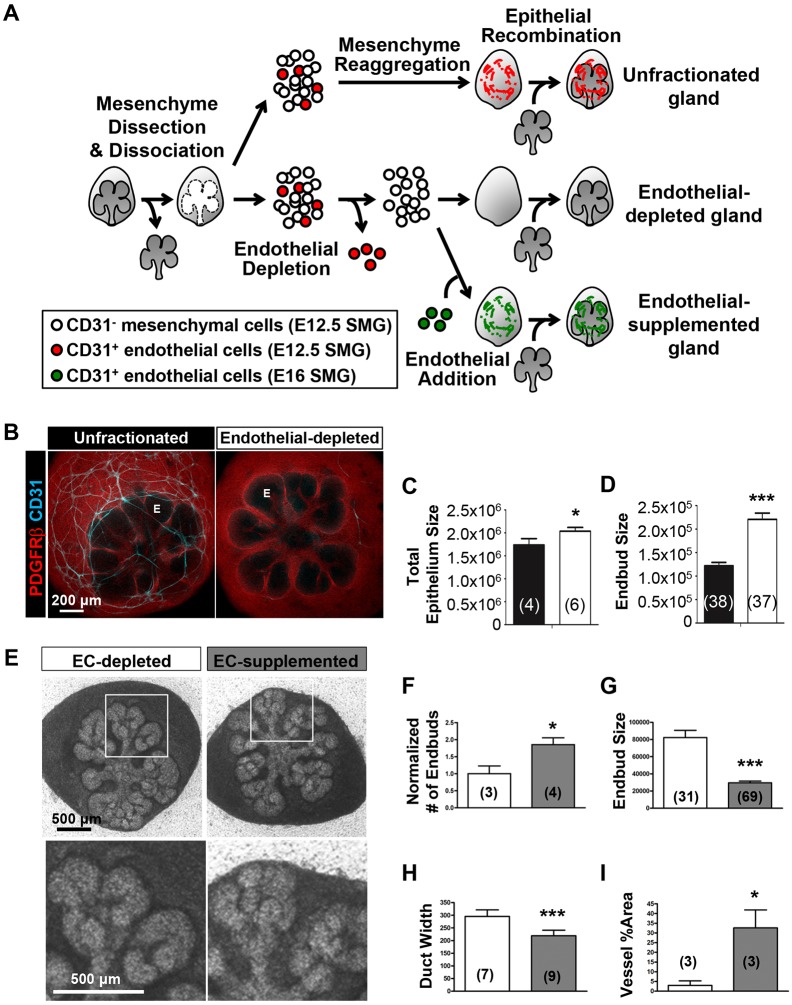
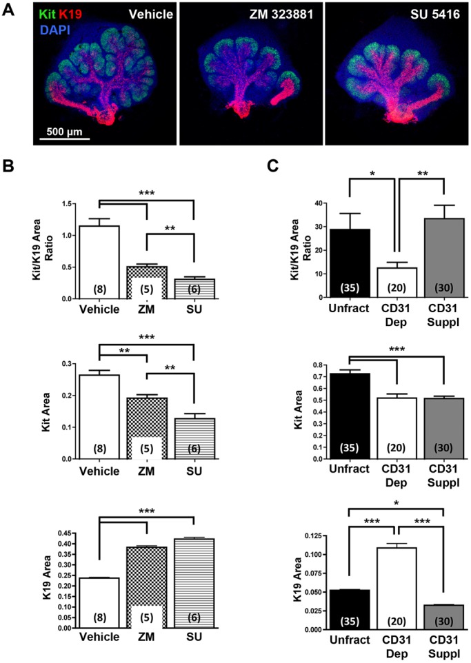
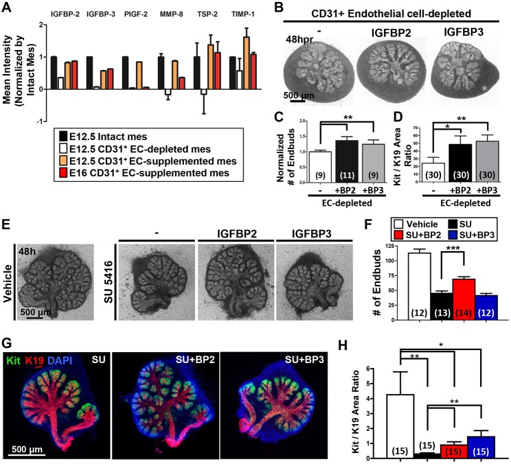
Perfusion-independent regulation of epithelial pattern formation by the vasculature during organ development and regeneration is of considerable interest for application in restoring organ function. During murine submandibular salivary gland development, the vasculature co-develops with the epithelium during branching morphogenesis; however, it is not known whether the vasculature has instructive effects on the epithelium. Using pharmacological inhibitors and siRNA knockdown in embryonic organ explants, we determined that VEGFR2-dependent signaling is required for salivary gland epithelial patterning. To test directly for a requirement for endothelial cells in instructive epithelial patterning, we developed a novel ex vivo cell fractionation/reconstitution assay. Immuno-depletion of CD31+ endothelial cells in this assay confirmed a requirement for endothelial cells in epithelial patterning of the gland. Depletion of endothelial cells or inhibition of VEGFR2 signaling in organ explants caused an aberrant increase in cells expressing the ductal proteins K19 and K7, with a reduction in Kit+ progenitor cells in the endbuds of reconstituted glands. Addition of exogenous endothelial cells to reconstituted glands restored epithelial patterning, as did supplementation with the endothelial cell-regulated mesenchymal factors IGFBP2 and IGFBP3. Our results demonstrate that endothelial cells promote expansion of Kit+ progenitor cells and suppress premature ductal differentiation in early developing embryonic submandibular salivary gland buds.

Keywords: Ductal differentiation; Endothelial cell; Endothelial cells; Epithelial patterning; Mouse; Progenitor cell; Salivary gland development.


Conflict of interest statement

Competing interests

The authors declare no competing or financial interests.

Figures

Fig. 1.
Fig. 1.
Co-development of epithelium and vasculature in early developing submandibular gland. (A) Mouse embryonic salivary glands were excised and subjected to ICC and confocal imaging to detect CD31+ (cyan) endothelial cells, shown superimposed on a brightfield image. (B) Single confocal images of the glands without brightfield are shown at higher magnification. A CD31+ semi-discontinuous vasculature with some isolated endothelial islands (white arrow) was observed in E11.5-E12.5 mesenchyme (M) that was largely isolated from the emerging epithelial bud (E). In E13-E13.5 glands, a largely continuous vessel network (yellow arrow) was observed that progressively surrounds the epithelium and is distinct from the developing nerves (Tubb3+, red). Note that the CD31+ vessels penetrate into the maturing epithelial clefts at E13 (white arrowhead). (C) CD31+ vessels persist in E12.5 glands cultured ex vivo (shown here after 6 h and after 24 h). CD31+ endothelial cells also express VEGFR2 (red) and collagen IV (green). Because collagen IV is incorporated into the basement membrane of both the endothelial cells and the epithelial cells, it defines the boundary between the epithelium and mesenchyme. CD31+/VEGFR2+/collagen IV+ endothelial cells are found adjacent to the initiating epithelial clefts at E12+6 h growth (arrowheads in upper panels) and ingress into maturing clefts after 24 h of culture (arrowheads in lower panels). (D) Schematic summarizing co-development of the primary epithelial bud with vasculature in the early submandibular gland. Endothelial cells comprising discontinuous vasculature are found in mesenchyme at E12/E12.5. Developing vessels subsequently undergo elongation and maturation concomitant with branching of the primary epithelial bud.
Fig. 2.
Fig. 2.
VEGFR2 signaling and vasculature development promote epithelial patterning in SMG organ culture. (A) Both pharmacological VEGFR2 inhibition (ZM 323881, 20 μM) and VEGFR2 siRNA (400 nM)-mediated knockdown inhibited vascular formation in E12.5 SMG organ explants cultured for 48 h, with reduced CD31+ (cyan) vasculature relative to negative controls, vehicle (DMSO) and non-targeting (NT) siRNA treatment. ICC and confocal microscopy were performed to outline the epithelial basement membrane (Col IV, green), mesenchyme (PDGFRβ, red) and vasculature (Col IV, green; CD31, cyan). Note that the epithelium appears to be less branched upon VEGFR2 inhibition or knockdown. E, epithelial bud; M, mesenchyme. (B) E13 glands were cultured as organ explants in the presence of two distinct VEGFR2 inhibitors (ZM 323881 and SU 5416) for 48 h at the indicated doses, again demonstrating disrupted epithelial organ patterning. (C-G) Brightfield images from multiple glands (n indicated on bars) were used to quantify epithelial structures. (C) Endbud numbers in VEGFR2-inhibitor treated glands were significantly reduced in ex vivo culture for 24 h and 48 h (n=total number of glands from four experiments). (D) The bud/duct ratio of treated glands, compared with vehicle control. Buds were reduced but ducts were enlarged (n=number of glands). In inhibitor-treated glands, individual bud size (E) was enlarged (n=total number of endbuds from five experiments) and the widths of secondary duct and primary duct (F,G) were widened, respectively (n=total number of ducts from five experiments). Data are mean±s.e.m. Two-way ANOVA (C) and Student's t-test (one-tailed) (D-G) were performed for statistical analysis (*P<0.05, **P<0.01, ***P<0.001).
Fig. 3.
Fig. 3.
CD31 cell-dependent vasculature development promotes epithelial patterning in an SMG cell fractionation/reconstitution assay. (A) SMG cell fractionation/reconstitution assay schematic. Unfractionated SMG mesenchyme amenable to cell immunodepletion was generated by microdissection of the mesenchyme from the epithelium followed by enzymatic dissociation of the mesenchyme to single cells and re-aggregation of the isolated mesenchymal cell population. Re-aggregated mesenchyme was then reconstituted with an intact microdissected E13 epithelial rudiment. For endothelial cell depletion, CD31+ endothelial cells were immunodepleted from fully dissociated mesenchyme cells using MACS with CD31 microbeads prior to re-aggregation of the dissociated mesenchyme and reconstitution with an intact epithelium. For endothelial cell supplementation, endothelial-depleted mesenchymal cells were mixed with MACS-isolated endothelial cells prior to re-aggregation of the mesenchyme and reconstitution with an intact epithelium. The reconstituted glands were cultured ex vivo for 48 h post-reconstitution. (B) Confocal images (maximum projection images) consistently showed a change in the epithelial patterning (no marker, black) with a mesenchymal marker (PDGFRβ in red) defining the mesenchymal shape. CD31+ vasculature (cyan) was present in unfractionated, but not in endothelial-depleted mesenchyme. E, endbud. (C,D) Epithelial area (C) (n=number of reconstituted glands) and endbud size (D) (n=number of endbuds from reconstituted glands shown in C) were enlarged in the absence of CD31+ endothelial cells. Black and white bars indicate unfractionated and endothelial-depleted glands, respectively. (E) Supplementation with E16 SMG-derived endothelial cells was performed with endothelial-depleted mesenchyme and epithelial recombination. Reconstituted glands were cultured for 48 h. Boxed areas are enlarged below. (F-H) Endothelial supplementation promoted epithelial branching (F) (n=number of reconstituted glands), smaller bud size (G) (n=number of endbuds in two reconstituted glands) and thinner ducts (H) (n=number of ducts in two reconstituted glands). (I) Increased vessel area was confirmed by CD31 staining in ICCs (n=number of reconstituted glands). In F-I, white and gray bars indicate endothelial-depleted (EC-depleted in E) and endothelial-supplemented (EC-supplemented in E), respectively. Data are mean±s.e.m. Student's t- test (one-tailed) was performed for statistical analysis (*P<0.05 ***P<0.001).
Fig. 4.
Fig. 4.
VEGFR signaling and vasculature development regulate SMG epithelial progenitor cell differentiation. (A) ICC and confocal microscopy show that pharmacological inhibition of VEGFR2 with ZM 323881 (20 µM) or SU 5416 (5 µM) expands the productal K19+ (red) cell population relative to the Kit+ (green) cell population in E12.5 SMGs organ explants grown for 48 h versus control glands. Nuclei were stained with DAPI (blue). (B) Maximum projection intensity images of the glands shown in A were used to quantify areas positive for Kit or K19 within the submandibular epithelium only. Oral epithelium at the base of the main duct was avoided to measure K19 expression area. The Kit+/K19+ ratio was decreased with both inhibitors, and Kit expression area was significantly decreased whereas K19 expression area was significantly increased. (C) In the SMG cell fractionation/reconstitution assay, similar CD31-dependent changes in SMG progenitor cells were also observed, with a marked increase in the K19+ productal cell population. Reconstituted glands were grown for 48 h. Total unfractionated dissociated mesenchyme (Unfract), endothelial depleted (CD31 Dep), endothelial supplementation after endothelial depletion (CD31 Suppl). ICC and confocal microscopy (single section of the middle of endbud) was performed to quantify Kit+ and K19+ expression areas (n=number of endbuds). Number of experiments: 7 unfractionated, 4 CD31-depleted and 6 CD31-supplemented. Endothelial cell immunodepletion significantly decreased the Kit+/K19+ ratio, which was partially rescued with endothelial supplementation and re-vascularization. Data are mean±s.e.m. Student's t-test was performed for statistical analysis (*P<0.05, **P<0.01, ***P<0.001).
Fig. 5.
Fig. 5.
Vascular endothelial cells influence soluble factors that regulate SMG epithelial patterning and differentiation. (A) Conditioned media collected from intact (black bar) or recombined mesenchyme, including endothelial depletion (white), E12.5 (orange) and E16 (red) CD31+ endothelial supplementation was analyzed with an angiogenesis proteome array. Select soluble factors abundance of which in the conditioned medias changed in response to CD31+ cell condition are shown, normalized to intact mesenchyme. Full normalized data for the array are available in Table S2. (B) Brightfield images of SMG cell glands reconstituted after CD31+ endothelial cell depletion cultured for 48 h without or with addition of recombinant IGFBP2 or IGFBP3 (1 μg/ml). (C,D) Quantitative analyses demonstrate that both IGFBP2 and IGFBP3 supplementation increased epithelial branching (C) (n=number of reconstituted glands) and the Kit+/K19+ area ratio (D) (n=total number of endbuds from six reconstituted glands). (E) Brightfield images of E13 SMG explants cultured ex vivo for 48 h in the absence or presence of 5 µM SU 5416 (SU), either with or without addition of recombinant IGFBP2 (BP2; 2 μg/ml) or IGFBP3 (BP3; 5 μg/ml). (F) Quantitative analysis of VEGFR2-inhibited glands shows disrupted epithelial patterning expressed as endbud numbers (three experiments). (G) ICC and confocal images representative of the partial rescue of Kit (green) relative to K19 (red) expression areas in the IGFBP2- and IGFBP3-supplemented, SU 5416-treated glands. (H) The Kit+/K19+ ratio was partially rescued with exogenous IGFBPs (n=total number of endbuds from three experiments). Data are mean±s.e.m. Student's t-test was performed for statistical analysis (*P<0.05, **P<0.01, ***P<0.001).

References

    1. An H. Y., Shin H. S., Choi J. S., Kim H. J., Lim J. Y. and Kim Y. M. (2015). Adipose mesenchymal stem cell secretome modulated in hypoxia for remodeling of radiation-induced salivary gland damage. PLoS ONE 10, e0141862 10.1371/journal.pone.0141862 - DOI - PMC - PubMed
    1. Azizoglu D. B. and Cleaver O. (2016). Blood vessel crosstalk during organogenesis-focus on pancreas and endothelial cells. Wiley Interdiscip. Rev. Dev. Biol. 5, 598-617. 10.1002/wdev.240 - DOI - PMC - PubMed
    1. Besnard V., Corroyer S., Trugnan G., Chadelat K., Nabeyrat E., Cazals V. and Clement A. (2001). Distinct patterns of insulin-like growth factor binding protein (IGFBP)-2 and IGFBP-3 expression in oxidant exposed lung epithelial cells. Biochim. Biophys. Acta 1538, 47-58. 10.1016/S0167-4889(00)00136-1 - DOI - PubMed
    1. Binkert C., Margot J. B., Landwehr J., Heinrich G. and Schwander J. (1992). Structure of the human insulin-like growth factor binding protein-2 gene. Mol. Endocrinol. 6, 826-836. - PubMed
    1. Bourner M. J., Busby W. H., Siegel N. R., Krivi G. G., McCusker R. H. and Clemmons D. R. (1992). Cloning and sequence determination of bovine insulin-like growth factor binding protein-2 (IGFBP-2) - comparison of its structural and functional properties with IGFBP-1. J. Cell. Biochem. 48, 215-226. 10.1002/jcb.240480212 - DOI - PubMed

Publication types

MeSH terms

Substances