. 2017 Jan 15;144(2):221-234.
doi: 10.1242/dev.130807.

Cartilage to bone transformation during fracture healing is coordinated by the invading vasculature and induction of the core pluripotency genes

Affiliations

Cartilage to bone transformation during fracture healing is coordinated by the invading vasculature and induction of the core pluripotency genes

Diane P Hu et al. Development. .

Abstract

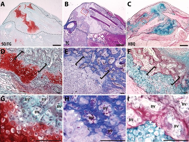
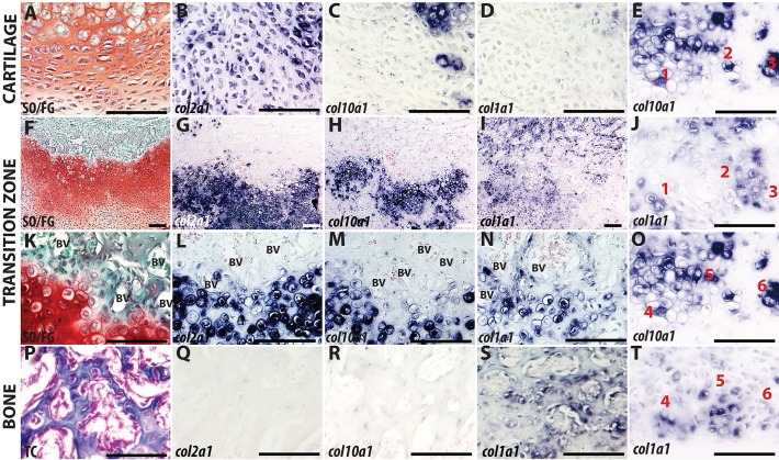
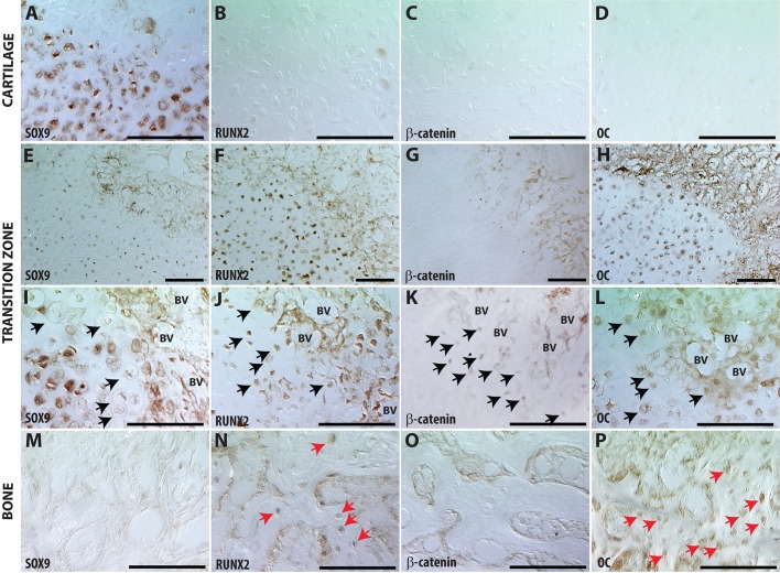
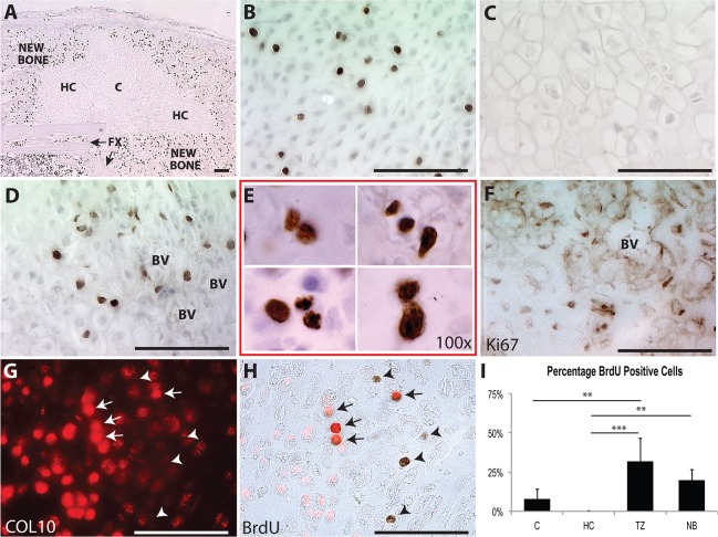
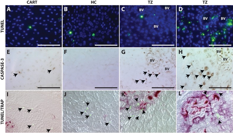
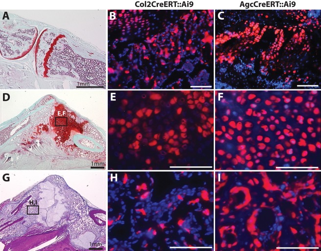
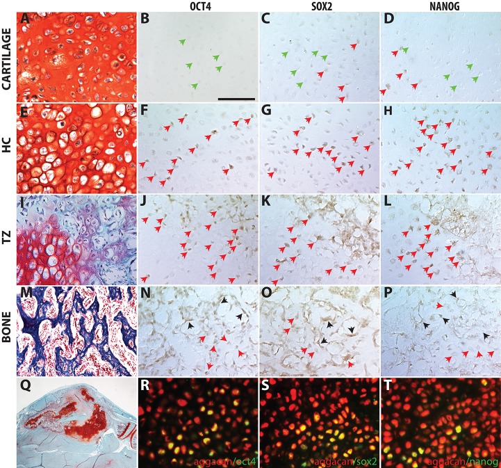
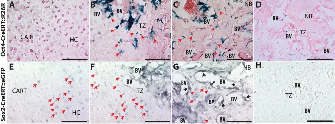
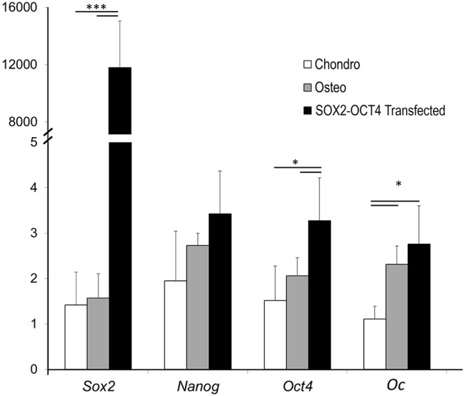
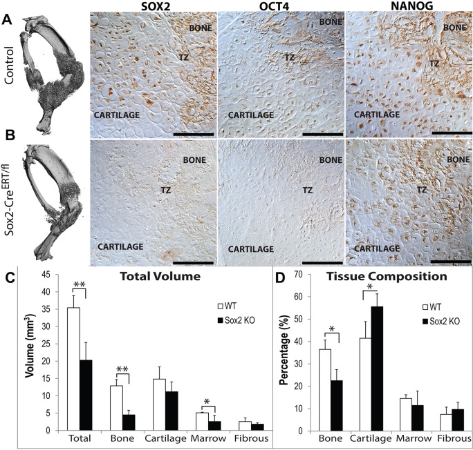
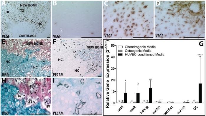
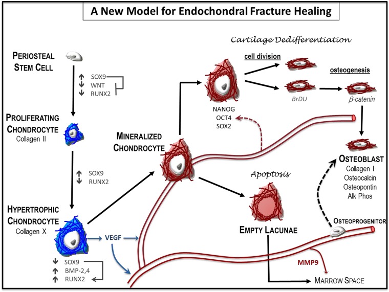
Fractures heal predominantly through the process of endochondral ossification. The classic model of endochondral ossification holds that chondrocytes mature to hypertrophy, undergo apoptosis and new bone forms by invading osteoprogenitors. However, recent data demonstrate that chondrocytes transdifferentiate to osteoblasts in the growth plate and during regeneration, yet the mechanism(s) regulating this process remain unknown. Here, we show a spatially-dependent phenotypic overlap between hypertrophic chondrocytes and osteoblasts at the chondro-osseous border in the fracture callus, in a region we define as the transition zone (TZ). Hypertrophic chondrocytes in the TZ activate expression of the pluripotency factors [Sox2, Oct4 (Pou5f1), Nanog], and conditional knock-out of Sox2 during fracture healing results in reduction of the fracture callus and a delay in conversion of cartilage to bone. The signal(s) triggering expression of the pluripotency genes are unknown, but we demonstrate that endothelial cell conditioned medium upregulates these genes in ex vivo fracture cultures, supporting histological evidence that transdifferentiation occurs adjacent to the vasculature. Elucidating the cellular and molecular mechanisms underlying fracture repair is important for understanding why some fractures fail to heal and for developing novel therapeutic interventions.

Keywords: Chondrocyte transformation; Endochondral ossification; Fracture repair; Pluripotency programs.


Conflict of interest statement

Competing interests

The authors declare no competing or financial interests.

Figures

Fig. 1.
Fig. 1.
Visualization of the chondro-osseous transition zone in a fracture callus. (A-C) Low magnification of a murine fracture callus, outlined with black dashed line, stained with (A) Safranin-O/Fast Green (SO/FG), (B) Modified Milligan's Trichrome (TC) or (C) Hall and Brunt Quadruple Stain (HBQ). (D-F) A magnified region of cartilage and bone from the fracture callus, outlined with a red box (A-C), with the TZ indicated by black brackets. (G-I) High magnification images of the TZ show the invading vasculature and the chondro-osseous junction. BV, blood vessel. Scale bars: 1 mm (A-C) and 100 µm (D-I).
Fig. 2.
Fig. 2.
Maturation of cartilage in the transition zone. Chondrocytes away from the TZ (A-D), compared with hypertrophic chondrocytes (HCs) in the TZ of murine fracture callus (E-O,T) or newly formed bone (P-S). Left column shows cartilage and bone histology stained with either SO/FG (A,F,K) or TC (P). In situ hybridization with Col2a1 (B,G,L,Q), Col10a1 (C,H,M,R) or Col1a1 (D,I,N,S). (E,J,O,T) Col10a1 and Col1a1 staining on adjacent sections 3-5 µm apart. Individual cells were tracked (cells 1-6) to demonstrate that staining does not overlap. Scale bars: 100 µm.
Fig. 3.
Fig. 3.
Hypertrophic chondrocytes adjacent to vasculature in the transition zone lose their chondrocyte phenotype and acquire an osteoblast phenotype. Immunohistochemistry in the cartilage away from the TZ (A-D), compared with HCs in the TZ (E-L) or new bone (NB) (M-P). (I-L) Black arrows indicate HCs in TZ that are Sox9 negative (I), and positive for Runx2 (J), β-catenin (K) or Oc (L). (M-P) Red arrows in NB tissue indicate Runx2+ (N) and Oc+ (P) cells. Scale bars: 100 µm.
Fig. 4.
Fig. 4.
Hypertrophic chondrocytes adjacent to vasculature in the transition zone re-enter the cell cycle. BrdU detection for entire fracture callus (A), immature chondrocytes away from the TZ (B), HCs away from the TZ (C), HCs in the TZ (D,E). (F,G) Ki67 and collagen X immunohistochemistry in HCs at the TZ. (H) Collagen X is observed to overlap with some of the BrdU+ cells (arrows), but not all (arrowheads). (I) Quantification of BrdU+/BrdU cells in fracture callus. Scale bars: 100 µm.
Fig. 5.
Fig. 5.
Cell death is not the predominant fate of hypertrophic chondrocytes during endochondral fracture repair. TUNEL staining (A-D), caspase-3 immunohistochemistry (E-H) or co-staining with TUNEL (green; I-L) to detect dying cells (black arrows) and TRAP to detect osteoclasts (purple/red; I-L) within immature chondrocytes (A,E,I), HCs away from the TZ (B,F,J), HCs within the TZ (C,G,K) and a region of maximal cell death at the TZ (D,H,L). Scale bars: 100 µm.
Fig. 6.
Fig. 6.
Chondrocytes give rise to osteoblasts and bone lining cells in the newly formed bone during fracture repair. Adult growth plate (A-C), cartilage (D-F) and newly formed bone (G-I) in fracture callus. Col2CreERT::Ai9 (B,E,H) or Agc1CreERT::Ai9 (C,F,I) mice 14 days post-fracture. Scale bars: 100 µm (B,C,E,F,H,I).
Fig. 7.
Fig. 7.
Expression of pluripotent stem cell programs in the transition zone of the fracture callus. Pluripotent stem cell protein is found in few cells at immature chondrocytes (A-D) but more frequently in HCs near the TZ (E-H) and within the TZ (I-L). (M-P) Within the NB, expression is observed in the HC encased in bone matrix and in bone-lining cells, but not in cells morphologically resembling osteoblasts/osteocytes. (Q) Low magnification of the representative fracture callus on which staining was performed on adjacent sections. (R-T) Cells from the chondrocyte lineage in Agc1CreERT::Ai9 mice (red) co-stained for Oct4 (R), Sox2 (S), Nanog (T) using an Alexa Fluor 488 secondary antibody (green); co-expression appears yellow. Green arrows indicate negative cells; red arrows, positive hypertrophic chondrocytes; black arrows, positive bone lining cells. Scale bar: 100 µm.
Fig. 8.
Fig. 8.
Transgenic reporter mice for Oct4 and Sox2 demonstrate transformation of chondrocytes into osteoblasts. Oct4-CreERT::R26R (A-D) and Sox2-CreERT::ROSAmT/mG (E-H) mice 14 days post-fracture in cartilage away from the TZ (A,D), TZ (B,E) and NB (C,F). (D) X-gal staining of C57B6. (H) Control with only secondary antibody. Red arrows indicate positive hypertrophic chondrocytes; black arrows, positive bone lining cells. Scale bars: 100 µm.
Fig. 9.
Fig. 9.
Transfection of OCT4 and SOX2 induces osteocalcin expression in fracture callus cartilage. Relative gene expression analysis of cartilage callus explants cultured in vitro in chondrogenic medium (white), osteogenic medium (gray), or chondrogenic medium with the OCT4-SOX2 transgene (black). Values are means±95% confidence. of n=6.
Fig. 10.
Fig. 10.
Conditional deletion of Sox2 compromises fracture healing. (A,B) µCT shows smaller callus size and immunohistomistry shows reduced expression of Sox2 and Oct4, but not Nanog. Tamoxifen was administered on days 4-7, 10 and 12 days post-fracture in (A) C57B6 control or (B) Sox2-CreER/fl mice. Histomorphometeric quantification of total tissue volumes (C) or tissue composition (D). Values are means±95% confidence of n=5. *P<0.05, **P<0.01. Scale bars: 100 µm.
Fig. 11.
Fig. 11.
Hypertrophic chondrocytes recruit vasculature through VEGF expression and vasculature coordinates activation of pluripotent programs. (A-D) Vegf immunohistochemistry (brown) of entire fracture callus (A), immature chondrocytes (B), HCs away from the TZ (C) or HCs in the TZ (D). HBQ (E,H) and Pecam1 (CD31) (F,I) immunohistochemistry (black) on adjacent sections. (G) Relative gene expression of pluripotent, chondrogenic and osteogenic genes for cartilage callus explants cultured in vitro with chondrogenic (white), osteogenic (gray), or HUVEC-CM (black). Values are means±95% confidence of n=4. *P<0.05, ***P<0.0005, ****P<0.0001. Scale bars: 100 µm.
Fig. 12.
Fig. 12.
A new model for endochondral ossification during fracture healing. Local osteochondral progenitors from the periosteum and endosteum are the stem cells that differentiate to form bone and cartilage in the fracture site. To generate the cartilage callus, osteochondral progenitors differentiate into chondrocytes (blue) that proliferate to generate the early soft callus. Chondrocytes within the callus mature to hypertrophy and express angiogenic factors that result in vascular invasion. Mineralization of the hypertrophic cartilage (red) occurs in the TZ where blood vessels have invaded. Some hypertrophic chondrocytes undergo cell death to facilitate remodeling of the solid cartilage callus and create marrow space within the trabecular bone. Other hypertrophic chondrocytes regain some stem cell-like properties by expressing the pluripotent transcription factors OCT4, SOX2 and NANOG. These large cells re-enter the cell cycle, divide and then transform into osteoblasts. The identity and origin of the signal that triggers the chondrocyte-to-osteoblast transformation remains unclear. Data from this paper and others suggest that both the vasculature and cell autonomous signals from the hypertrophic chondrocyte may facilitate this change (dotted arrows). This model does not exclude previously proposed systems in which osteoblasts in the newly formed bone are derived from osteoprogenitors that are brought in by the invading vasculature.

References

    1. Arnold K., Sarkar A., Yram M. A., Polo J. M., Bronson R., Sengupta S., Seandel M., Geijsen N. and Hochedlinger K. (2011). Sox2(+) adult stem and progenitor cells are important for tissue regeneration and survival of mice. Cell Stem Cell 9, 317-329. 10.1016/j.stem.2011.09.001 - DOI - PMC - PubMed
    1. Aspenberg P., Genant H. K., Johansson T., Nino A. J., See K., Krohn K., García-Hernández P. A., Recknor C. P., Einhorn T. A., Dalsky G. P. et al. (2010). Teriparatide for acceleration of fracture repair in humans: a prospective, randomized, double-blind study of 102 postmenopausal women with distal radial fractures. J. Bone Miner. Res. 25, 404-414. 10.1359/jbmr.090731 - DOI - PubMed
    1. Bahney C. S., Hu D. P., Taylor A. J., Ferro F., Britz H. M., Hallgrimsson B., Johnstone B., Miclau T. and Marcucio R. S. (2014). Stem cell-derived endochondral cartilage stimulates bone healing by tissue transformation. J. Bone Miner. Res. 29, 1269-1282. 10.1002/jbmr.2148 - DOI - PMC - PubMed
    1. Bahney C. S., Hu D. P., Miclau T. III and Marcucio R. S. (2015). The multifaceted role of the vasculature in endochondral fracture repair. Front. Endocrinol. 6, 4 10.3389/fendo.2015.00004 - DOI - PMC - PubMed
    1. Bais M., McLean J., Sebastiani P., Young M., Wigner N., Smith T., Kotton D. N., Einhorn T. A. and Gerstenfeld L. C. (2009). Transcriptional analysis of fracture healing and the induction of embryonic stem cell-related genes. PLoS ONE 4, e5393 10.1371/journal.pone.0005393 - DOI - PMC - PubMed

Publication types

MeSH terms