. 2017 Feb 15;144(4):635-648.
doi: 10.1242/dev.140855. Epub 2017 Jan 17.

Efficient CRISPR/Cas9-assisted gene targeting enables rapid and precise genetic manipulation of mammalian neural stem cells

Affiliations

Efficient CRISPR/Cas9-assisted gene targeting enables rapid and precise genetic manipulation of mammalian neural stem cells

Raul Bardini Bressan et al. Development. .

Abstract

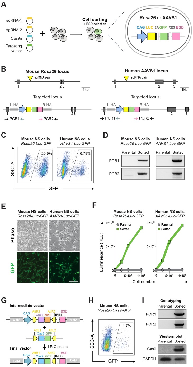
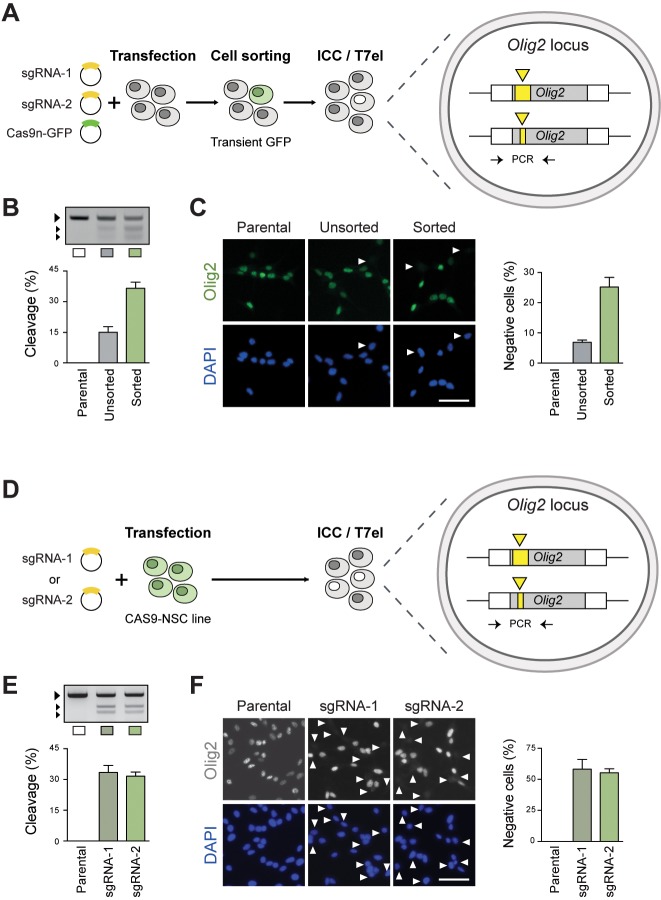
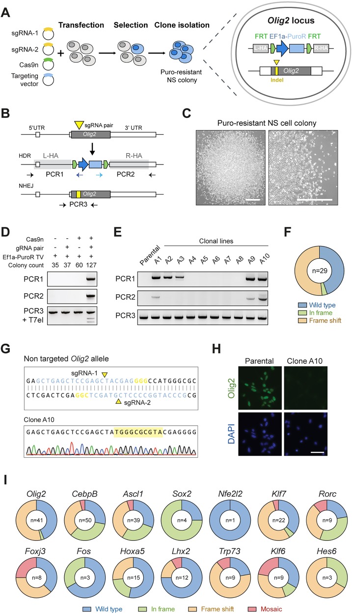
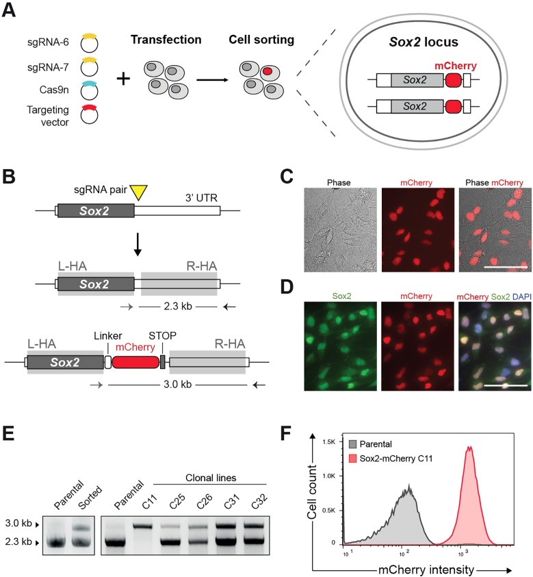
Mammalian neural stem cell (NSC) lines provide a tractable model for discovery across stem cell and developmental biology, regenerative medicine and neuroscience. They can be derived from foetal or adult germinal tissues and continuously propagated in vitro as adherent monolayers. NSCs are clonally expandable, genetically stable, and easily transfectable - experimental attributes compatible with targeted genetic manipulations. However, gene targeting, which is crucial for functional studies of embryonic stem cells, has not been exploited to date in NSC lines. Here, we deploy CRISPR/Cas9 technology to demonstrate a variety of sophisticated genetic modifications via gene targeting in both mouse and human NSC lines, including: (1) efficient targeted transgene insertion at safe harbour loci (Rosa26 and AAVS1); (2) biallelic knockout of neurodevelopmental transcription factor genes; (3) simple knock-in of epitope tags and fluorescent reporters (e.g. Sox2-V5 and Sox2-mCherry); and (4) engineering of glioma mutations (TP53 deletion; H3F3A point mutations). These resources and optimised methods enable facile and scalable genome editing in mammalian NSCs, providing significant new opportunities for functional genetic analysis.

Keywords: CRISPR/Cas9; Epitope tagging; Gene targeting; Genome editing; Glioblastoma; Homologous recombination; Neural stem cell; Transcription factor.


Conflict of interest statement

Competing interests

The authors declare no competing or financial interests.

Figures

Fig. 1.
Fig. 1.
Mouse and human NSCs are amenable to CRISPR/Cas9-mediated gene targeting. (A) Schematic of the experimental strategy for generating constitutive Luciferase (Luc)-GFP expressing mouse and human NSCs by gene targeting at the safe harbour loci Rosa26 or AAVS1. Targeting vectors contained the CAG-Luc-2A-GFP tethered by an IRES to a blasticidin resistance cassette (BSD); the expression cassettes were flanked by ∼1 kb long homology arms. (B) Schematic depiction of the targeting strategy for the Rosa26 (left) and AAVS1 loci (right). Exons are shown as dark grey blocks. Light grey rectangles indicate the location of the homology arms flanking the expression cassette (L-HA, left homology arm; R-HA, right homology arm). CRISPR sgRNA target sites are indicated with yellow triangles. Horizontal arrows indicate genotyping PCR primers used to confirm on-target integration of the expression cassette. (C) Using FACS, targeted cells were enriched on the basis of GFP expression after blastidicin selection. Wild-type non-transfected mouse and human NSCs were used as a control to set gates for cell sorting. SSC, side scatter. (D) PCR-based genotyping using primer sets 1 and 2 (depicted in A) confirmed correct targeted integration of the CAG-Luc-2A-GFP cassette into the Rosa26 and AAVS1 loci. Non-transfected parental cells were used as negative control for the genotyping. (E) Representative live phase contrast and wide-field fluorescence microscopy images of sorted GFP-positive mouse and human NSCs. Scale bar: 100 μm. (F) Luciferase levels were determined using a microplate reader and confirmed functionality of the targeted cassette in both mouse and human cells. (G) Schematic of the Gateway cloning-based strategy for repurposing targeting vectors with different cassettes of interest. In the example shown, the Luc-2A-GFP in the Rosa26 targeting vector is replaced by a Cas9-GFP expression cassette via LR Gateway cloning. (H) Mouse NSCs were transfected with the Rosa26 Cas9-GFP targeting vector and enriched by FACS on the basis of GFP expression. (I) PCR-based genotyping (top) confirms correct insertion of Cas9-GFP expression cassette at the Rosa26 locus; western immunoblotting (bottom) confirms high levels of Cas9 protein expression in a clonal NSC line derived from the GFP-sorted cells.
Fig. 2.
Fig. 2.
Olig2 knockout in mouse NSCs via CRISPR/Cas9-induced NHEJ repair. (A) Experimental strategy for Olig2 deletion in wild-type (WT) mouse NSCs using transient plasmid delivery. Cells were transiently transfected with the CRISPR sgRNA pair (target site shown as yellow triangles) together with a Cas9n-2A-GFP plasmid. Transfected cells were enriched by FACS on the basis of GFP expression. (B) Generation of indel mutations in the transfected cells was assessed using a T7EI cleavage assay. Larger arrow indicates the predicted WT/uncleaved PCR product; smaller arrows indicate T7EI-cleaved fragments used to estimate indel frequency. (C) ICC was used to determine the frequency of Olig2-negative cells, which result from biallelic frameshift mutations (white arrowheads). Graph shows percentage of Olig2-negative cells relative to total DAPI-stained nuclei. Scale bar: 50 µm. (D,E) Experimental strategy for Olig2 deletion in the CAS9 NSC line. Cells were transfected with the two CRISPR sgRNAs individually (D) and cleavage at target site confirmed by the T7E1 assay (E). (F) Efficiency of biallelic knockout was quantified by ICC for Olig2. Scale bar: 50 µm.
Fig. 3.
Fig. 3.
CRISPR/Cas9-mediated homologous recombination enables facile knockout of transcription factor genes in mouse NSCs. (A) Schematic of the experimental strategy to knock out Olig2 via CRISPR/Cas9-assisted gene targeting. Targeted cells are enriched by puromycin selection and should emerge as discrete NSC colonies. Biallelic knockout clones are expected to have one allele replaced by the Ef1α-PuroR cassette and the second allele damaged by an indel mutation (yellow rectangle) at the CRISPR sgRNA cutting site (yellow triangle). (B) Representation of the mouse Olig2 locus (top), and predicted targeted alleles following HDR (middle) or NHEJ (bottom). Olig2 coding sequence is shown in dark grey. Adjacent white rectangles represent untranslated regions (5′ and 3′ UTR). PCR genotyping (PCR1 and 2, left and right arms, respectively) were designed with primers within the Ef1α-PuroR cassette and outside of the homology arms (light grey rectangles). PCR3 product could be used in a T7EI assay or Sanger sequencing to confirm the presence of indels in the NHEJ allele. (C) Representative phase contrast image of exemplar puromycin-resistant NSC colony, which emerged after transfection with Olig2 targeting vector. Scale bars: 200 µm. (D) PCR genotyping of pooled puromycin-resistant colonies after transfection with Olig2 targeting vector (Ef1α-PuroR TV) alone or in combination with CRISPR Cas9 nickase and/or sgRNA pair. PCR1 and PCR2 were used to confirm correct HR event at Olig2 locus (top and middle); T7EI assay for checking NHEJ-mediated damaged at the sgRNA targeted site (bottom). (E) Representative genotyping PCR results of ten puromycin-resistant clones, picked and expanded as clonal lines following transfection with targeting vector and CRISPR/Cas9 components. (F) Summary of types of indel mutations identified by Sanger sequencing of PCR3 product (shown in B) in correctly targeted Olig2 clones. Wild type, no indel; in frame, 3N; frame shift, 3N+1 or 3N+2. (G) Representative Sanger sequencing trace of one Olig2 targeted clone showing 10 bp insertion within the remaining Olig2 coding exon. (H) ICC in wild-type parental cells and Olig2 clone harbouring the 10 bp insertion confirms complete ablation of the Olig2 protein. Scale bar: 50 µm. (I) Scale-up of the same strategy used for Olig2 in 14 other transcription factors and frequency of indel types present on the second allele of correctly targeted clones. Mosaics contained more than one indel within the same clone and were identified by mixed Sanger sequencing traces (see Fig. S5B).
Fig. 4.
Fig. 4.
Generation of homozygous Sox2-mCherry reporter NSC line. (A) Schematic of the experimental strategy for knock-in of mCherry reporter at the Sox2 C terminus. Cells were transfected with the targeting vector together with sgRNA pair and Cas9n plasmids. After 10 days, mCherry-positive cells were isolated by cell sorting. (B) Representation of Sox2 targeted locus and PCR-based genotyping. No promoter sequence is contained within the targeting vector (promoterless construct) and therefore mCherry expression is expected only when the Sox2 locus is correctly targeted. sgRNA pair was designed to cut in the 3′ UTR close to the stop codon. mCherry was fused to the Sox2 coding sequence through a flexible peptide linker (white box). (C) Live images of sorted cells showing nuclear localised mCherry signal. Scale bar: 50 µm. (D) ICC reveals overlap of mCherry and Sox2 in the nucleus. Scale bar: 50 µm. (E) PCR genotyping of clonal lines derived after cell sorting. Homozygous clone (C11) shows only an upper band (3.0 kb), whereas heterozygously targeted clones also show lower, wild-type band (2.3 kb). (F) Flow cytometry histogram confirming consistent mCherry expression in homozygous Sox2-mCherry clonal line. Parental NSCs (grey line) were used to set the gates.
Fig. 5.
Fig. 5.
CRISPR/Cas9-based gene targeting enables epitope tagging of endogenous transcription factor genes. (A) Representation of strategy to knock in the V5 tag in-frame into the Olig2 C terminus using a single-stranded oligonucleotide DNA (ssODN) as a donor template. V5 tag sequence is shown in green, homology arms in grey and stop codon in black. Yellow triangle represents the sgRNA target site (sgRNA-3) at Olig2 3′UTR. (B) Schematic depicting the three strategies employed for the knock-in of the V5 epitope tag: (a) delivery of plasmids encoding sgRNA and Cas9-2A-GFP followed by FACS enrichment; (b) delivery of a ribonucleoprotein complex made by conjugating in vitro transcribed (IVT) sgRNA and recombinant Cas9 protein (rCas9); (c) delivery of IVT sgRNA into the CAS9 NSCs (constitute Cas9 expressing from Rosa26). (C) Representative image of V5-tagged Olig2 cells (arrowheads) identified using ICC against the V5 tag. DAPI was used for nuclear staining. Scale bar: 50 µm. (D) Quantification of Olig2-V5-positive cells using the three delivery strategies (shown in B). Values represent the percentage of V5-positive cells relative to total DAPI nuclear counting. (E) PCR-based genotyping of representative v5-Olig2 clones derived from the bulk cells transfected with rCas9+IVT sgRNA complex. PCR1 used primers within the V5 sequence and outside the R-HA. Homozygosity was confirmed using primers flanking the V5 insertion site (PCR2). Homozygous targeted clones (A1 and A4) were identified by a single upper band 42 bp higher than the control, WT band. Clones displaying two bands were considered as heterozygous. (F) ICC for the V5 tag and Olig2 in homozygously tagged clonal lines. As anticipated, V5 staining is nuclear localised and overlaps with native Olig2 staining. Scale bar: 50 µm.
Fig. 6.
Fig. 6.
Delivery of glioma-relevant mutations into genetically normal human NSCs. (A) Schematic depiction of the targeting strategy to knock out TP53 via gene targeting. CRISPR sgRNA pair targeting site is indicated with a yellow triangle. Horizontal arrows indicate PCR genotyping primers for assaying TP53 locus targeting (PCR1 and PCR2) and presence of an indel within the second allele (PCR3). (B) Representative phase contrast images of puromycin-resistant human NSC colony after 10 days of selection. Right-hand image is a magnification of the boxed area on the left. Scale bar: 100 µm. (C) PCR-based genotyping (top) and WB analysis (bottom) of human TP53 targeted NSC clonal lines. Parental, non-transfected cells were used as a control. (D) Sanger sequencing trace of an exemplar correctly targeted clone harbouring an 85 bp deletion on the second allele. (E) Growth curve analysis of the TP53 targeted clones harbouring the 85 bp deletion confirming the positive proliferative effect of p53 ablation. (F) Illustration of the strategy for gene targeting the first H3F3A coding exon. Yellow triangle indicates sgRNA pair targeting site. Horizontal arrows indicate PCR genotyping primers. (G) ICC using a V5 tag-specific antibody to identify targeted cells (arrowheads). DAPI was used for nuclear staining. Scale bar: 50 µm. (H) Western blotting using V5 tag and histone H3-specific antibodies and PCR genotyping of parental and transfected cells. (I) Sanger sequencing traces of the genotyping PCR products confirm presence of the point mutations in the V5 tag-positive cells.

References

    1. Bernstein B. E., Mikkelsen T. S., Xie X., Kamal M., Huebert D. J., Cuff J., Fry B., Meissner A., Wernig M., Plath K. et al. (2006). A bivalent chromatin structure marks key developmental genes in embryonic stem cells. Cell 125, 315-326. 10.1016/j.cell.2006.02.041 - DOI - PubMed
    1. Brennan C. W., Verhaak R. G. W., McKenna A., Campos B., Noushmehr H., Salama S. R., Zheng S., Chakravarty D., Sanborn J. Z., Berman S. H. et al. (2013). The somatic genomic landscape of glioblastoma. Cell 155, 462-477. 10.1016/j.cell.2013.09.034 - DOI - PMC - PubMed
    1. Bruggeman S. W. M., Hulsman D., Tanger E., Buckle T., Blom M., Zevenhoven J., van Tellingen O. and van Lohuizen M. (2007). Bmi1 controls tumor development in an Ink4a/Arf-independent manner in a mouse model for glioma. Cancer Cell 12, 328-341. 10.1016/j.ccr.2007.08.032 - DOI - PubMed
    1. Campos P. B., Sartore R. C., Abdalla S. N. and Rehen S. K. (2009). Chromosomal spread preparation of human embryonic stem cells for karyotyping. J. Vis. Exp. 4, 1512 10.3791/1512. - DOI - PMC - PubMed
    1. Capecchi M. R. (2005). Gene targeting in mice: functional analysis of the mammalian genome for the twenty-first century. Nat. Rev. Genet. 6, 507-512. 10.1038/nrg1619 - DOI - PubMed

Publication types

MeSH terms