

Review
doi: 10.1101/cshperspect.a031336.
Toward the Atomic Structure of PrPSc
Affiliations
- PMID: 28096267
- PMCID: PMC5585850
- DOI: 10.1101/cshperspect.a031336
Item in Clipboard
Review
Toward the Atomic Structure of PrPSc
Cold Spring Harb Perspect Biol.
.
Abstract
In this review, we detail our current knowledge of PrPSc structure on the basis of structural and computational studies. We discuss the progress toward an atomic resolution description of PrPSc and results from the broader field of amyloid studies that may further inform our knowledge of this structure. Moreover, we summarize work that investigates the role of PrPSc structure in its toxicity, transmissibility, and species specificity. We look forward to an atomic model of PrPSc, which is expected to bring diagnostics and/or therapeutics to the field of prion disease.
Copyright © 2017 Cold Spring Harbor Laboratory Press; all rights reserved.
Figures
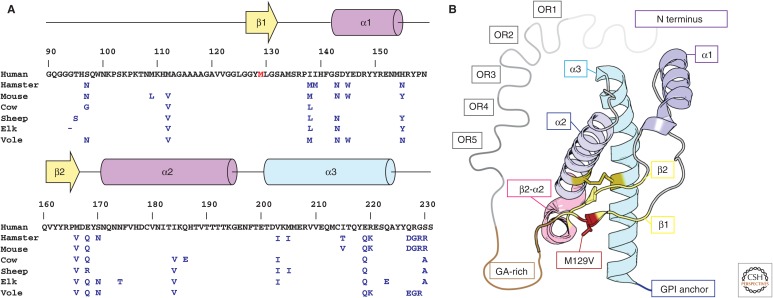
Sequence and structure of human PrPC. (A) Primary sequence and secondary structure of human PrPC residues 90–230. Residues that differ between hamster, mouse, cow, sheep, elk, and bank vole sequences are indicated in blue, and those of the native human sequence are shown in black. The genetic variant M129V is shown in red. (B) Model of the crystal structure of the M129V variant of human PrPC. Secondary structure elements are labeled using a color scheme that follows A; these include five octapeptide repeats (OR1–5, gray), a glycine-alanine-rich stretch (GA-rich, brown), two β-strands (β1, β2, yellow), and three α-helices (α1–α3, magenta-blue), variant M129V (red), the β2–α2 turn (pink), and a C-terminal glycosylphosphatidylinositol (GPI) anchor (blue).
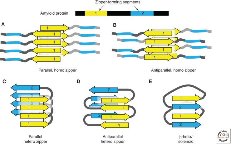
Steric zipper hypothesis of amyloid fibril structure. Amyloid proteins often contain two or more amyloidogenic segments (steric zippers). The homo or hetero association of zipper segments results in five types of β-sheet architecture, giving rise to a large variety of possible assemblies of amyloid molecules. (A,B) The homotypic association between identical zipper segments in parallel (A) and antiparallel (B) arrangement. (C,D) The hetero association between different zipper segments in parallel (C) and antiparallel (D) arrangement. (E) “Hetero” interaction is built from the parallel stacking of different segments in the right-hand β-helix or solenoid.
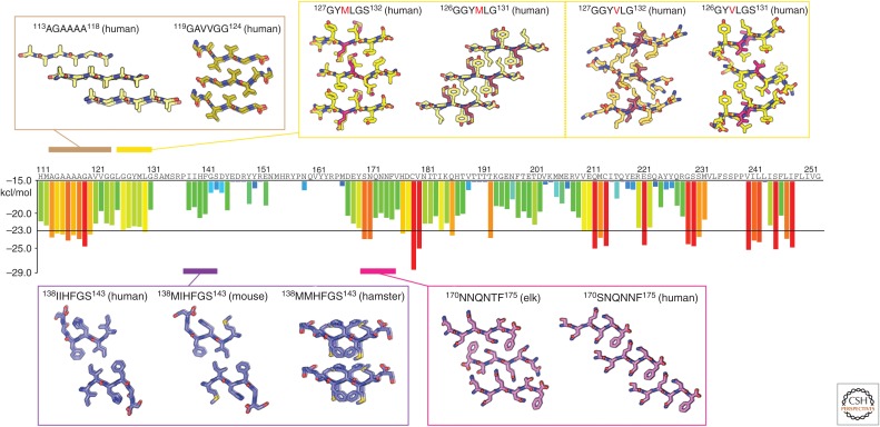
Atomic structures of PrP steric zippers. The propensity for any given six-residue segment in the sequence of human PrP to form a steric zipper was estimated using the ZipperDB database (http://services.mbi.ucla.edu/zipperdb/ ). The evaluation of a single six-residue segment gives an energy score shown in kcal/mol on the bar graph axis (left). A colored bar representing each score is assigned to the first residue of each six-residue window throughout the PrP sequence. More negative scores are more favorable, and segments with a score of less than −23 kcal/mol are the most likely to form zippers. Atomic structures of zipper-forming segments from four regions of the sequence are shown. Two structures of the human sequence cover the region residues 113–124. Several structures cover residues 126–132 and contain either a methionine or valine at position 129. Structures from human, mouse, and hamster sequences span residues 138–143. Two structures—one with an elk sequence and the other human—span residues 170–175. As indicated in Table 1, numbers correspond to the human sequence.
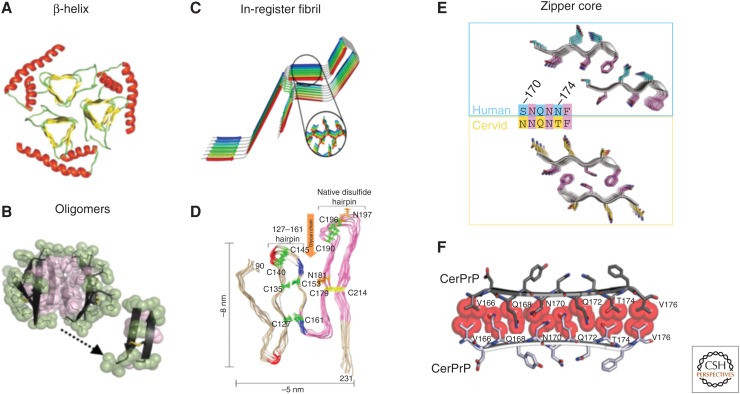
Proposed models for PrPSc. (A) A triangular β-helix model of PrPSc was based on a transmission electron microscopy (TEM) image. (Courtesy of Govaerts et al. 2004; reprinted, with permission, from the National Academy of Sciences, U.S.A. © 2001.) (B) An atomic structure for a PrP oligomer was determined from pairs of naturally disulfide-linked segments with PrP sequences. (Courtesy of Apostol et al. 2013; reprinted, with permission, from the American Chemical Society © 2013.) (C,D) In-register fibril models have been obtained from different experiments. (C) An in-register stacking model was proposed based on electron paramagnetic resonance (EPR) of human PrP fibrils converted in vitro. (Courtesy of Cobb et al. 2007; reprinted, with permission, from the National Academy of Sciences, U.S.A. © 2007.) (D) Another parallel in-register β-sheet architecture using solid-state NMR spectroscopy of recombinant PrP amyloid fibrils that were seeded with PrPSc. (Courtesy of Groveman et al. 2014; adapted, with permission, © 2014, by the American Society for Biochemistry and Molecular Biology.) (E,F) Zipper structures of segments in or near the β2–α2 loop highlight its importance in conversion and transmission. (E) Microcrystal structures of human (cyan, top) and cervid (yellow, lower) β2–α2 loop segments belong to different classes of steric zippers, supporting the idea that side-chain mismatches may account for species barriers in prion disease. (Courtesy of Apostol et al. 2011; adapted, with permission, © 2011 American Chemical Society.) (F) Zipper model of the β2–α2 loop highlights tight, complementary side-chain interactions that are required for efficient prion conversion. (Courtesy of Kurt et al. 2014b; adapted, with permission, © 2014, by the American Society for Biochemistry and Molecular Biology.)
References
Publication types
MeSH terms
Substances
LinkOut - more resources
Full Text Sources
Other Literature Sources
Research Materials