. 2017 May;28(5):1603-1613.
doi: 10.1681/ASN.2016030343. Epub 2017 Jan 17.

Anti-Factor B and Anti-C3b Autoantibodies in C3 Glomerulopathy and Ig-Associated Membranoproliferative GN

Affiliations

Anti-Factor B and Anti-C3b Autoantibodies in C3 Glomerulopathy and Ig-Associated Membranoproliferative GN

Maria Chiara Marinozzi et al. J Am Soc Nephrol. 2017 May.

Abstract

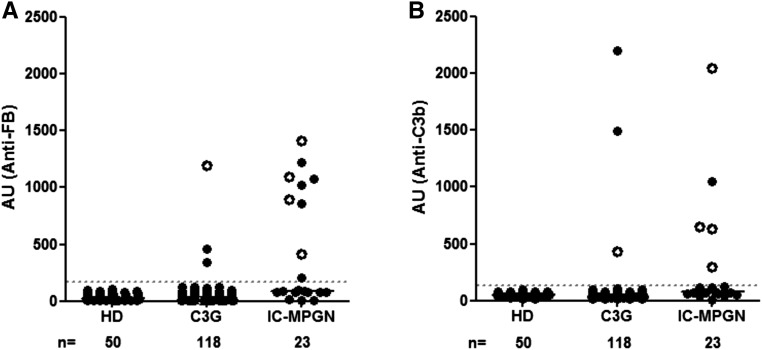
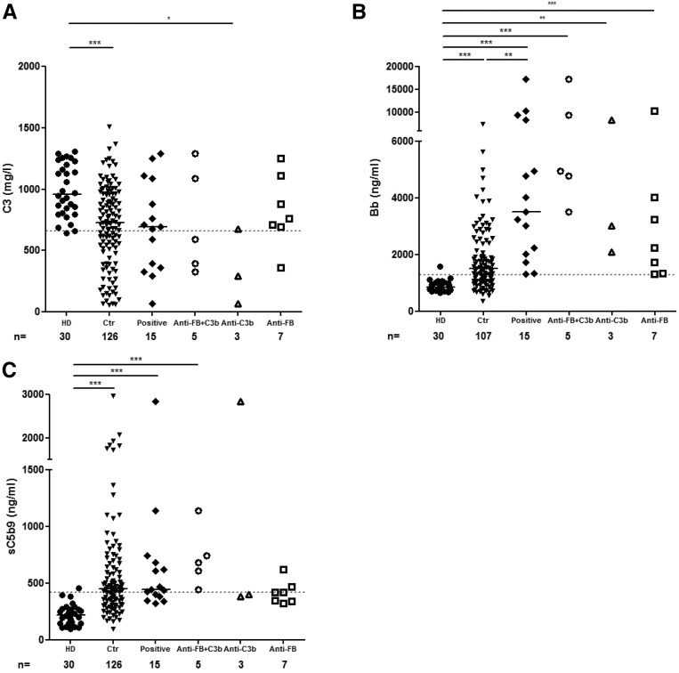
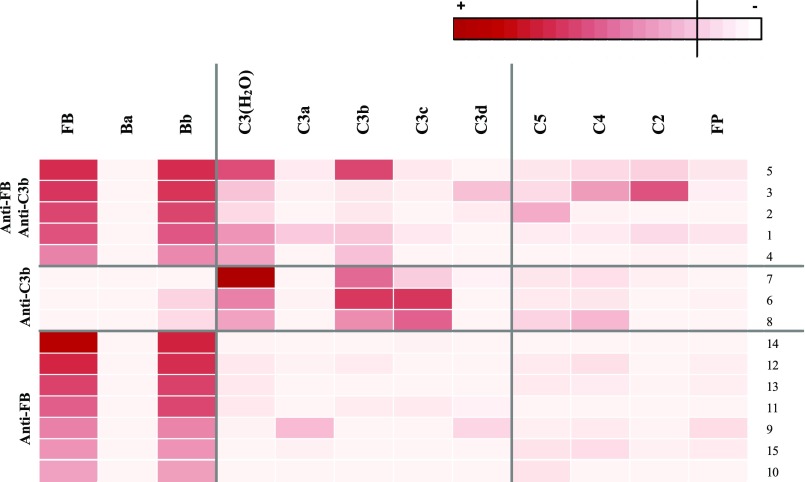
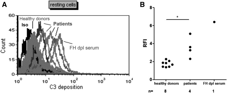
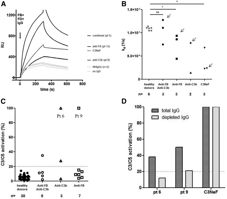
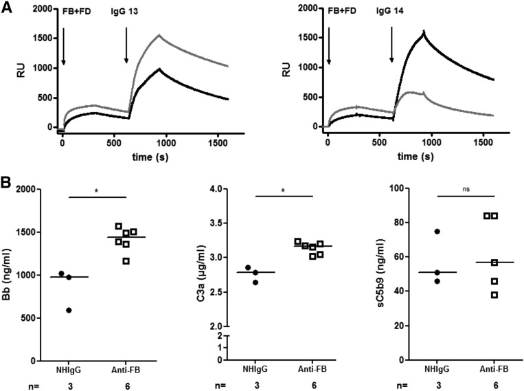
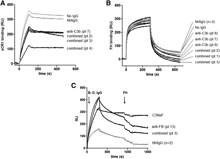
In C3 glomerulopathy (C3G), the alternative pathway of complement is frequently overactivated by autoantibodies that stabilize the C3 convertase C3bBb. Anti-C3b and anti-factor B (anti-FB) IgG have been reported in three patients with C3G. We screened a cohort of 141 patients with C3G and Ig-associated membranoproliferative GN (Ig-MPGN) for anti-FB and anti-C3b autoantibodies using ELISA. We identified seven patients with anti-FB IgG, three patients with anti-C3b IgG, and five patients with anti-FB and anti-C3b IgG. Of these 15 patients, ten were diagnosed with Ig-MPGN. Among those patients with available data, 92% had a nephrotic syndrome, 64% had AKI, and 67% had a documented infection. Patients negative for anti-C3b and anti-FB IgG had much lower rates of infection (17 [25%] patients with C3G and one [10%] patient with Ig-MPGN). After 48 months, four of 15 (26%) positive patients had developed ESRD or died. All 15 patients had high plasma Bb levels, six (40%) patients had low levels of C3, and nine (60%) patients had high levels of soluble C5b9. In vitro, IgG purified from patients with anti-FB Abs selectively enhanced C3 convertase activity; IgG from patients with anti-C3b/anti-FB Abs enhanced C3 and C5 cleavage. IgG from patients with anti-C3b Abs stabilized C3bBb and perturbed C3b binding to complement receptor 1 but did not perturb binding to factor H. In conclusion, the prevalence of anti-C3b/anti-FB Abs and alternative pathway activation is similar in Ig-MPGN and C3G, suggesting similar pathogenic mechanisms. Identification of the underlying defect in Ig-MPGN could lead to improved treatment.

Keywords: chronic glomerulonephritis; clinical immunology; complement.


Figures

Figure 1.
Figure 1.
Detection of anti-FB and anti-C3b autoantibodies. Plasma samples from patients with C3G (n=118) and Ig-MPGN (n=23) were tested. Anti-FB and anti-C3b Abs were expressed in AUs. White circles represent samples with double positivity. The cutoff of positivity was determined with plasma samples from 50 healthy donors (HDs).
Figure 2.
Figure 2.
Biopsies on native kidney from two patients. Patients with (A1–A3) isolated anti-FB (patient 12) and (B1–B3) isolated anti-C3b (patient 9). (A1 and B1) Trichrome coloration (×40), (A2 and B2) C3 staining (×40), and (A3 and B3) C5b9 staining (×40).
Figure 3.
Figure 3.
Complement activation biomarker assessments. Plasma levels of (A) C3, (B) Bb, and (C) sC5b9 of 15 anti-C3b/anti-FB–positive plasma samples and 30 healthy controls. *P<0.05; **P>0.01; ***P>0.001. Ctr, control cohort of C3G without anti-C3b and anti-FB autoantibodies; HD, healthy donor.
Figure 4.
Figure 4.
Epitope mapping of the anti-FB and anti-C3b IgG. Heat map showing the binding levels of patient IgG to the different antigens. The intensity of the binding is represented by red shading. FP, factor properdine.
Figure 5.
Figure 5.
C3 deposition on endothelial cells. (A) Histogram of the C3 deposition on the surface of resting endothelial cells incubated with the sera from patients 3, 9, 12, and 14, which was measured by flow cytometry. Three serum samples from healthy donors served as controls. FH-depleted serum was used as a positive control for AP dysregulation. A representative histogram is shown. (B) Quantification of the C3 deposition on resting endothelial cells. *P<0.05. iso, isotype; dpl, depleted; RFI, ratio fluorescency/intensity.
Figure 6.
Figure 6.
Effects of positive patient IgG on the C3/C5 convertase of the AP. (A) Sensorograms illustrating real-time C3 convertase in the presence of positive patient IgG. After coupling C3b on the biosensor chip, we flowed a mix of FB, factor D, and IgG across it. The sensorograms of the IgG of one patient from each group (combined anti-FB/anti-C3b–, anti-FB–, or anti-C3b–positive patients) and two IgG from healthy donors (NHIgGs) and in the absence of IgG were reported as examples. C3NeF-positive IgG was used as a positive control. IgG from patient 5 (combined anti-FB/anti-C3b Ab), patient 13 (anti-C3b), and patient 8 (anti-FB) enhanced the formation of the C3 convertase. FD, factor D. (B) Calculated off rate of the C3 convertase formed in the presence of patient IgG. A lower off rate than controls signals an increased stabilization of the complexes. The effect of the IgG seemed to be related to the stabilization of the convertase, because the off rate of the triple complex is lower in the presence of IgG from our patients compared with IgG from healthy donors. Arrows indicate the Kd relative to the sensorograms shown in A. (C) Effect of positive IgG samples on the C3/C5 convertase stabilization. C3 convertase was assembled on the surface of sheep erythrocytes by incubating C3b-bound sheep erythrocytes with FB, factor D, and patient IgG. The addition of rat serum triggered lysis of cells, in which C5 lytic sites were still present after spontaneous decay. The results are expressed in percentages of residual C3/C5 convertase sites. (D) Effect on the C3/C5 activation of the depletion of anti-C3b and anti-FB Ab from two positive samples (patients 6 and 9). Depleted anti-C3b and anti-FB Ab IgG from IgG positive reduced the percentage of the C3/C5 convertases compared with total IgG, showing that they are responsible for this activity. The C3NeF sample, used as a positive control and treated as the anti-C3b IgG, maintained full activity after treatment. NH, normal human; Pt, patient; RU, response unit. P<0.05.
Figure 7.
Figure 7.
Overall functional effect of the anti-FB–positive IgG. (A) Sensorograms illustrating real-time interaction between two examples of positive anti-FB IgG (patients 13 and 14) with C3(H2O)Bb (gray line) or C3bBb convertase (black line). Patient IgG is able to bind to its target, even when it is complexed in the convertase. (B) Complement activation after addition of purified positive anti-FB IgG to normal human serum or IgG from healthy donors (NHIgG). Release of C3a, Bb, and sC5b9. Statistical analyses were performed by GraphPad software using a Mann Whitney test. *P<0.05. FD, factor D; NH, normal human; RU, response unit.
Figure 8.
Figure 8.
The anti-C3b autoantibodies decreased the binding of the regulator CR1 but did not decrease the binding of FH to C3b. (A) Binding of CR1 to C3b pre-exposed to patient IgG. (B) Binding of FH to C3b pre-exposed to patient IgG. (C) Dissociation of the C3 convertase formed in presence of patient IgG by FH. IgG from a C3NeF-positive patient served as a positive control for a convertase, resistant to decay by FH. Anti-C3b–positive IgG is in black, IgG from healthy donors is in light gray, and dissociation in the absence of IgG is shown as a dashed line. IgG from a C3NeF-positive patient, stabilizing the convertase and making it resistant to FH decay, is shown as a dark dashed line. Pt, patient; RU, response unit.

References

    1. Merle NS, Noe R, Halbwachs-Mecarelli L, Fremeaux-Bacchi V, Roumenina LT: Complement system part II: Role in immunity. Front Immunol 6: 257, 2015 - PMC - PubMed
    1. Merle NS, Church SE, Fremeaux-Bacchi V, Roumenina LT: Complement system part I - molecular mechanisms of activation and regulation. Front Immunol 6: 262, 2015 - PMC - PubMed
    1. Zipfel PF, Skerka C, Chen Q, Wiech T, Goodship T, Johnson S, Fremeaux-Bacchi V, Nester C, de Córdoba SR, Noris M, Pickering M, Smith R: The role of complement in C3 glomerulopathy. Mol Immunol 67: 21–30, 2015 - PubMed
    1. Servais A, Noël LH, Roumenina LT, Le Quintrec M, Ngo S, Dragon-Durey MA, Macher MA, Zuber J, Karras A, Provot F, Moulin B, Grünfeld JP, Niaudet P, Lesavre P, Frémeaux-Bacchi V: Acquired and genetic complement abnormalities play a critical role in dense deposit disease and other C3 glomerulopathies. Kidney Int 82: 454–464, 2012 - PubMed
    1. Cook HT, Pickering MC: Histopathology of MPGN and C3 glomerulopathies. Nat Rev Nephrol 11: 14–22, 2015 - PubMed