. 2017 Jan 31;114(5):E781-E790.
doi: 10.1073/pnas.1613422114. Epub 2017 Jan 17.

Pathogen-mediated manipulation of arthropod microbiota to promote infection

Affiliations

Pathogen-mediated manipulation of arthropod microbiota to promote infection

Nabil M Abraham et al. Proc Natl Acad Sci U S A. .

Abstract

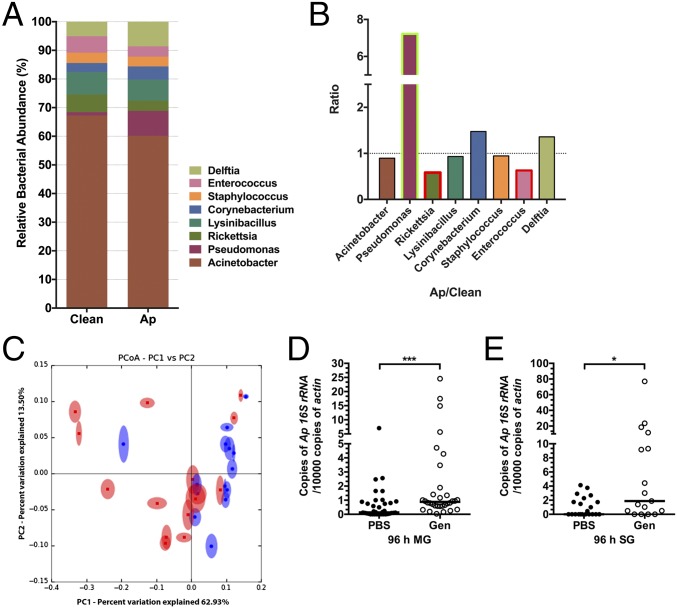
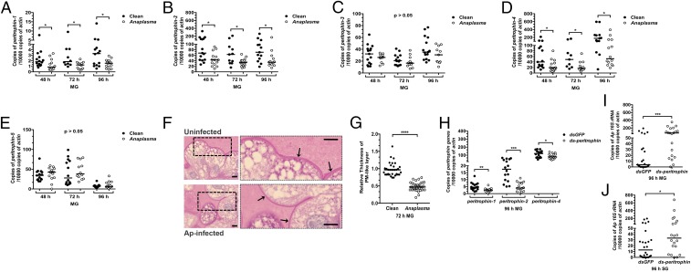
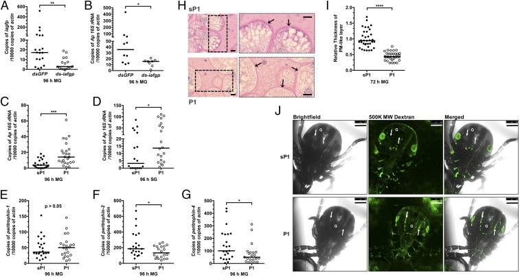
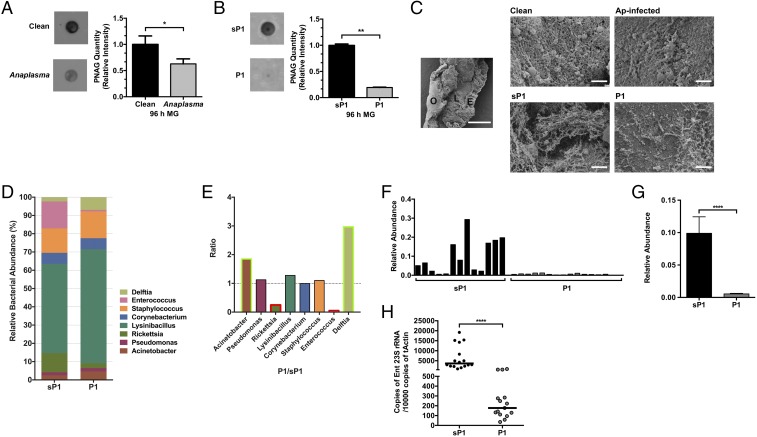
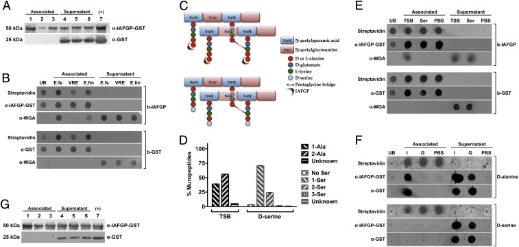
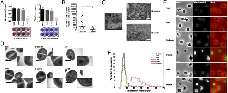
Arthropods transmit diverse infectious agents; however, the ways microbes influence their vector to enhance colonization are poorly understood. Ixodes scapularis ticks harbor numerous human pathogens, including Anaplasma phagocytophilum, the agent of human granulocytic anaplasmosis. We now demonstrate that A. phagocytophilum modifies the I. scapularis microbiota to more efficiently infect the tick. A. phagocytophilum induces ticks to express Ixodes scapularis antifreeze glycoprotein (iafgp), which encodes a protein with several properties, including the ability to alter bacterial biofilm formation. IAFGP thereby perturbs the tick gut microbiota, which influences the integrity of the peritrophic matrix and gut barrier-critical obstacles for Anaplasma colonization. Mechanistically, IAFGP binds the terminal d-alanine residue of the pentapeptide chain of bacterial peptidoglycan, resulting in altered permeability and the capacity of bacteria to form biofilms. These data elucidate the molecular mechanisms by which a human pathogen appropriates an arthropod antibacterial protein to alter the gut microbiota and more effectively colonize the vector.

Keywords: Anaplasma; Ixodes scapularis; antifreeze protein; biofilm; microbiome.


Conflict of interest statement

The authors declare no conflict of interest.

Figures

Fig. 1.
Fig. 1.
A. phagocytophilum changes the tick microbiota and dysbiosis enhances A. phagocytophilum colonization. (A and B) Comparison of the gut microbial composition of uninfected and A. phagocytophilum-infected fed nymphs. (A) Total bacterial abundance and (B) ratio (Ap/Clean), at the taxonomic rank of genus, of uninfected (Clean) and A. phagocytophilum-infected (Ap) fed nymphs. Green and red box outlines indicate the genera with increased and decreased bacterial abundance in A. phagocytophilum-infected nymphs. (C) Principal coordinate analysis (PCoA) of the microbial communities from uninfected (blue) and A. phagocytophilum-infected (red) fed nymphs based on weighted UniFrac. Statistical significance was calculated using ANOSIM method, P = 0.028. (D and E) The qRT-PCR assessment of the A. phagocytophilum burden in normal (PBS) and dysbiosed gentamicin-treated (Gen) nymphal tick (D) guts (MG) and (E) salivary glands (SG). Horizontal bars represent the median. Results were pooled from three independent experiments. Statistical significance was calculated using a two-tailed nonparametric Mann−Whitney test (***P < 0.001; *P < 0.05).
Fig. 2.
Fig. 2.
The PM influences colonization of the tick by A. phagocytophilum. (AE) A. phagocytophilum influences the expression of (A) peritrophin-1, (B) peritrophin-2, (C) peritrophin-3, (D) peritrophin-4, and (E) peritrophin-5 in the tick gut (MG) as assessed by qRT-PCR. Results were pooled from three independent experiments, and statistical significance was calculated using a two-tailed nonparametric Mann−Whitney test (*P < 0.05). (F) PAS staining of Carnoy’s fixed and sectioned fed guts from uninfected and A. phagocytophilum-infected nymphs. Boxed outlines within the images on the left have been magnified 2.5×. Arrows indicate the PAS-positive PM-like layer. (Scale bar, 10 μm.) (G) Quantification of the relative thickness of the PM-like layer from uninfected and A. phagocytophilum-infected nymphal guts (MG). (HJ) The qRT-PCR examination of the expression of (H) peritrophin-1, peritrophin-2, and peritrophin-4 upon peritrophin dsRNA mixture injection of nymphal ticks, and qRT-PCR expression of the A. phagocytophilum burden in (I) guts (MG) and (J) salivary glands of dsgfp- and ds-peritrophin-injected nymphs fed on A. phagocytophilum-infected mice. Each dot represents one nymph. Horizontal bars represent the median. Statistical significance was calculated from three independent experiments using a two-tailed nonparametric Mann−Whitney test (****P < 0.0001; ***P < 0.001; **P < 0.01; *P < 0.05).
Fig. 3.
Fig. 3.
IAFGP and its peptide derivative, P1, influence the PM and A. phagocytophilum colonization of tick gut. (A and B) The qRT-PCR examination of the expression of (A) iafgp and (B) the A. phagocytophilum burden in dsgfp- and ds-iafgp-injected nymphs [gut (MG)] fed on A. phagocytophilum-infected mice. Each dot represents one nymph. (C and D) The qRT-PCR assessment of the A. phagocytophilum burden in the (C) guts (MG) and (D) salivary glands with P1- or control [scrambled P1(sP1)]-injected nymphs fed on A. phagocytophilum-infected mice. Each dot represents one nymph. (EG) The qRT-PCR analysis of expression levels of (E) peritrophin-1, (F) peritrophin-2, and (G) peritophin-4 in P1- and sP1-injected nymphs [gut )MG)]. Each dot represents one nymph. (H) PAS staining of Carnoy’s fixed and sectioned fed guts from P1- and sP1-injected nymphs. Arrows indicate the PAS-positive PM-like layer. Boxed outlines within the images on the left have been magnified 2×. (Scale bar, 10 μm.) (I) Quantification of relative thickness of the PM-like layer from P1- and sP1-injected nymphal guts (MG). Statistical significance was calculated using a two-tailed nonparametric Mann−Whitney test from three pooled experiments (****P < 0.0001; ***P < 0.001; **P < 0.01; *P < 0.05). (J) P1 injection of ticks improves the permeability of the PM. Confocal microscopy of 24-h P1- or sP1-injected nymphs that were capillary-fed Fluorescein-conjugated 500,000 MW dextran (500K MW Dextran). Magnification is 10×. G marks the gut diverticula within the tick. The arrows point to the hemocoel outside the gut. (Scale bar, 250 μm.)
Fig. 4.
Fig. 4.
IAFGP and P1 alter biofilm formation and microbiota in ticks. (A and B) The relative amount of PNAG extracted from (A) uninfected and. A. phagocytophilum-infected nymphs and (B) P1- vs. sP1-injected nymphs and their respective quantification. Relative intensity of PNAG blots were analyzed by ImageJ (graphs). Graphical data were pooled from two independent experiments, and statistical significance was calculated using a two-tailed nonparametric Mann−Whitney test (**P < 0.01; *P < 0.05). (C) SEM of tick gut biofilm from uninfected (Clean) and A. phagocytophilum (Ap) infected nymphs (Top) and sP1- vs. P1-injected nymphs (Bottom). Representative images are shown for all samples. A representative uninfected (Clean) sample is provided (Left, Middle) at low magnification (500×) to visualize a dissected gut revealing the outside of the gut (O), epithelial cells lining the gut (E), and luminal content (L) (scale bar, 50 μm). Representative images displayed were taken at high magnification (∼8,500×; scale bar, 2 μm) focusing on the luminal content. Ap-infected and P1-injected guts show looser biofilms without dense connecting fibers or matrix-like material associated with reduced biofilms. Clean and sP1 guts, contrastingly, have thicker and well-connected matrix like material with embedded bacteria (cocci) representing robust biofilms. (D and E) Genus level (D) total bacterial composition of sP1- and P1-injected nymphs and (E) fold changes (P1/sP1) of major genera. Green and red box outlines represent genera with increased or decreased abundance in P1-injected nymphs. Statistical significance was calculated using a two-tailed nonparametric Mann−Whitney test (****P < 0.0001). (FH) Comparison of the levels of Enterococcus species within the tick gut microbiota between sP1- and P1-injected nymphal ticks. (F and G) Relative abundance of Enterococci (F) among individual samples and (G) across all samples (pooled). (H) P1-injected nymphs show reduced levels of Enterococci as determined by qRT-PCR using Enterococci specific primers compared with sP1-injected ticks.
Fig. 5.
Fig. 5.
IAFGP binds to the terminal d-alanine residue of Enterococcus sp. and S. aureus peptidoglycan. (A) E. faecalis (lanes 1 and 4), E. faecium (lanes 2 and 5), and vancomycin-resistant E. faecalis (lanes 3 and 6) bacteria were incubated with either recombinant GST-IAFGP or GST alone. Bound (Associated) and unbound (Supernatant) protein was detected by immunoblot. Recombinant GST-IAFGP or GST alone (lane 7) was used as a positive control. (B) IAFGP displays varied binding to Enterococcal peptidoglycan. Streptavidin-coated magnetic Dynabeads bound to biotinylated IAFGP-GST (b-IAFGP) or GST alone (b-GST) were incubated with peptidoglycan muropeptides extracted from E. faecalis (E.fs), vancomycin-resistant E. faecalis (VRE), and E. faecium (E.fm) cells grown in BHIG. Biotinylated proteins bound to beads were detected using an infrared-labeled streptavidin probe. IAFGP-GST was detected using a polyclonal murine primary antibody, and GST was detected using a monoclonal murine primary antibody. Muropeptides from cells were detected using a polyclonal WGA antibody. Unbound protein (UB) was also collected and spotted. Associated or Supernatant fractions represent the fractions that were either pulled down with the magnetic bead or remained unbound. (C) Schematic describing S. aureus peptidoglycan and binding site of IAFGP. The S. aureus peptidoglycan backbone is a polymer consisting of glycan strands of alternating sugar subunits: NAG and NAM. The NAM subunits have short peptides (muropeptides) comprising l-alanine, d-glutamate, l-lysine, and two d-alanines. The cross-link between neighboring muropeptides is mediated by a transpeptidase enzyme and allows for the formation of a short peptide interbridge consisting of five glycines (pentaglycine bridge). IAFGP is hypothesized to bind to the terminal d-alanine residue of the stem pentapeptide (Top). Growth of S. aureus in an alternative amino acid like d-serine (Bottom) replaces the terminal d-alanine residue with the newly supplied d-serine. (D) Muropeptide composition of S. aureus peptidoglycan. S. aureus grown in either TSB or medium supplemented with 125 mM d-serine were analyzed for their muropeptide residue composition. Data represent the percentage of total bacterial muropeptides that incorporate a given residue(s) at its carboxyl terminus. “No Ser” represents d-alanine terminating muropeptides. Data were pooled from three independent experiments ± SEM. (E and F) IAFGP binds to the terminal d-alanine residue of the S. aureus muropeptide chain. (E) Streptavidin-coated magnetic Dynabeads bound to b-IAFGP or b-GST were incubated with muropeptides obtained from cells grown in either TSB or medium supplemented with 125 mM d-serine. (F) Experiment was similarly performed as in E using synthetic biotinylated pentapeptides containing either alanine (d-alanine) or serine (d-serine) residues bound to beads and incubated with IAFGP-GST (I) or GST alone (G). PBS was used as a negative binding control. Associated or Supernatant fractions represent the fractions that were either pulled down with the magnetic bead or remained unbound. (G) IAFGP does not bind WTA. Wildtype S. aureus strain SA113 (lanes 1 and 3) and its isogenic WTA-deficient mutant SA113 ΔtagO (lanes 2 and 5) and complemented strain SA113 pRBtagO (lanes 3 and 7) were tested for binding to recombinant GST-IAFGP or control GST alone. Bound (Associated) and unbound (Supernatant) protein was detected by immunoblot analysis. Recombinant GST-IAFGP or GST alone (lane 7) without any bacteria was used as a positive control.
Fig. 6.
Fig. 6.
d-serine incorporation causes alterations to bacterial biofilms and peptidoglycan. (A) Biofilm formation of S. aureus SA113 (Left) and E. faecalis MMH594 (Right) cultures was determined in respective media. Dissolved stains were pooled from three independent experiments ± SEM; representative images of the biofilm plates with two representative wells per condition are shown below the graphs. Statistical significance was calculated using one-way ANOVA with Dunnett’s multiple comparison posttest (***P < 0.0001). (B) d-serine-injected nymphal ticks show reduced levels of Enterococci compared with control d-alanine-injected ticks, as determined by qRT-PCR using Enterococci specific primers. Statistical significance was calculated using a two-tailed nonparametric Mann−Whitney test (****P < 0.0001; ***P = 0.0003). (C) SEM of tick gut biofilms from d-alanine- versus d-serine-injected nymphal ticks. d-alanine-treated representative sample taken at low magnification (500×) is provided to visualize a dissected gut revealing the outside of the gut (O), epithelial cells lining the gut (E), and luminal content (L). (Scale bar, 50 μm.) Representative images are shown for both samples taken at high magnification (∼8,500×; scale bar, 2 μm) focusing on the luminal content. Compared with dense matrix biofilms with d-alanine treatment, there is a severe scarcity of the biofilm-associated polymeric matrix and bacteria with the d-serine treatment. (D) Overnight cultures of S. aureus SA113 in TSB medium alone or medium supplemented with 125 mM d-alanine, 125 mM d-serine, 0.1 mg/mL GST-IAFGP, or 0.1 mg/mL GST alone were processed for transmission electron microscopy. Representative images are shown. Cultures were also grown in an equivalent volume of PBS (buffer control). (Left) Representative images were taken at 60,000 magnification. (Scale bar, 500 nm.) Inset images were taken at 150,000 magnification (box length = scale bar = 800 nm). (E) Bacterial cells cultured in TSB or medium supplemented with PBS, d-alanine, d-serine, GST, or IAFGP were stained with the NAG binding analog Wheat Germ Agglutinin–Texas Red and imaged by phase-contrast and epifluorescence microscopy. (Scale bar, 2 μm.) (F) Population analysis, at the single-cell level, demonstrates that IAFGP and d-serine treatments result in an increase in fluorescent signal intensity. Total fluorescent signal intensities were normalized by cell area, and histograms were depicted as a function of population frequency; n > 500 cells for each treatment was pooled from two independent experiments.

References

    1. Goddard J. Infectious Diseases and Arthropods. Humana; Totowa, NJ: 2000.
    1. Hill CA, Kafatos FC, Stansfield SK, Collins FH. Arthropod-borne diseases: Vector control in the genomics era. Nat Rev Microbiol. 2005;3(3):262–268. - PubMed
    1. Andreotti R, et al. Assessment of bacterial diversity in the cattle tick Rhipicephalus (Boophilus) microplus through tag-encoded pyrosequencing. BMC Microbiol. 2011;11(1):6. - PMC - PubMed
    1. Boissière A, et al. Midgut microbiota of the malaria mosquito vector Anopheles gambiae and interactions with Plasmodium falciparum infection. PLoS Pathog. 2012;8(5):e1002742. - PMC - PubMed
    1. Broderick NA, Lemaitre B. Gut-associated microbes of Drosophila melanogaster. Gut Microbes. 2012;3(4):307–321. - PMC - PubMed

Publication types