. 2017 Apr;23(4):578-585.
doi: 10.1261/rna.060186.116. Epub 2017 Jan 17.

Structural basis for guanidine sensing by the ykkC family of riboswitches

Affiliations

Structural basis for guanidine sensing by the ykkC family of riboswitches

Robert A Battaglia et al. RNA. 2017 Apr.

Abstract

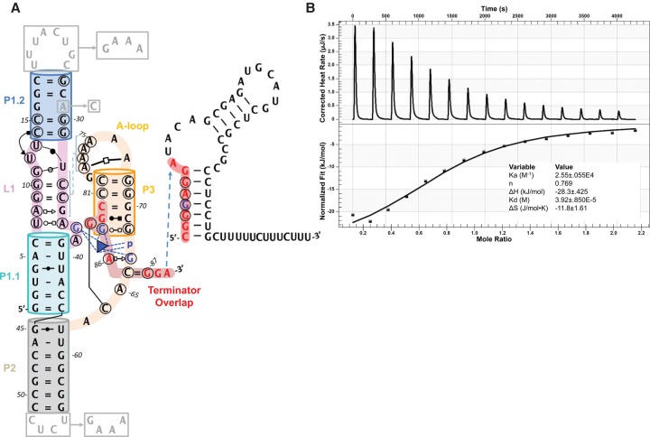
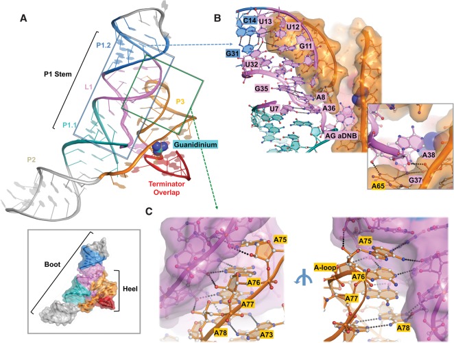
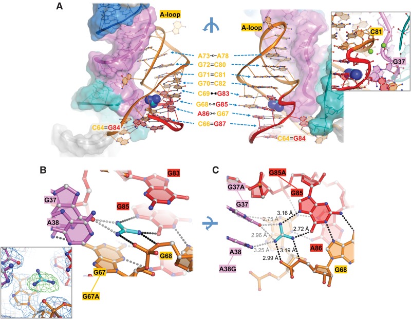
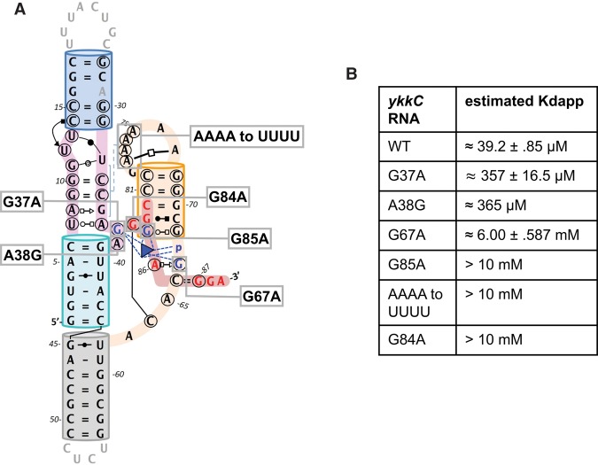
Regulation of gene expression by cis-encoded riboswitches is a prevalent theme in bacteria. Of the hundreds of riboswitch families identified, the majority of them remain as orphans, without a clear ligand assignment. The ykkC orphan family was recently characterized as guanidine-sensing riboswitches. Herein we present a 2.3 Å crystal structure of the guanidine-bound ykkC riboswitch from Dickeya dadantii The riboswitch folds into a boot-shaped structure, with a coaxially stacked P1/P2 stem forming the boot, and a 3'-P3 stem-loop forming the heel. Sophisticated base-pairing and cross-helix tertiary contacts give rise to the ligand-binding pocket between the boot and the heel. The guanidine is recognized in its positively charged guanidinium form, in its sp2 hybridization state, through a network of coplanar hydrogen bonds and by a cation-π stacking contact on top of a conserved guanosine residue. Disruption of these contacts resulted in severe guanidinium-binding defects. These results provide the structural basis for specific guanidine sensing by ykkC riboswitches and pave the way for a deeper understanding of guanidine detoxification-a previously unappreciated aspect of bacterial physiology.

Keywords: Dickeya dadantii; RNA structure; gene regulation; guanidine; orphan riboswitch.


Figures

FIGURE 1.
FIGURE 1.
Secondary structure model and ITC binding curve of Dda_ykkC. (A) 2D-representation of Dda_ykkC with Leontis–Westhof notation describing base interactions observed in the crystal structure. P1.1 (teal), L1 (violet), P1.2 (marine), P2 (gray), and P3 (orange) are shown. Circled residues are 97% conserved in subtype 1 ykkC riboswitches. Residues and phosphate contacting guanidinium (blue triangle) are highlighted in blue. Red residues participate in alternative terminator stem. Gray residues, boxes, and arrows show changes to WT sequence in the crystal construct. (B) ITC analysis of guanidinium binding by WT Dda_ykkC.
FIGURE 2.
FIGURE 2.
Overall structure of Dda_ykkC and detailed cross-helix interactions. (A) Cartoon representation of guanidinium (cyan) bound Dda_ykkC. Surface representation showing boot-like shape (bottom inset). (B) Detailed non-WC contacts in L1 region including AG aDNB and cross-helix contacts (inset). (C) Minor groove interaction between P3 A-loop and L1. Ribose zipper contacts are shown from the front (left) and base-specific contacts from the rear (right).
FIGURE 3.
FIGURE 3.
Guanidinium recognition in the binding pocket. (A) Front (right) and rear (left) views of Dda_ykkC P3 stem. Base pairs are detailed with Leontis–Westhof notation in the middle with arrows indicating the location of the pair in front and rear views. Inset shows magnesium ions coordinated by the P1 and P3 backbones. (B) Side view of the binding pocket showing cation–π stacking interaction between guanidinium (cyan) and G67 (orange). Inset shows omit-map electron density of the binding pocket at 1.5 σ generated by simulated annealing refinement. Black dashes represent hydrogen bonds. Gray dashes represent weak electrostatic interactions. (C) Top view of the binding pocket showing hydrogen-bonding network. Mutants designed for binding affinity experiments are connected by lines to their corresponding residue labels. Interatomic distances are given next to dashes.
FIGURE 4.
FIGURE 4.
Structure-guided mutagenesis evaluated by ITC analysis. (A) 2D-representation of Dda_ykkC showing the positions with which each mutant corresponds. (B) Table displaying estimates of WT and mutant binding affinities for guanidinium.

References

    1. Adams PD, Afonine PV, Bunkóczi G, Chen VB, Davis IW, Echols N, Headd JJ, Hung L-W, Kapral GJ, Grosse-Kunstleve RW, et al. 2010. PHENIX: a comprehensive Python-based system for macromolecular structure solution. Acta Crystallogr Sect D Biol Crystallogr 66: 213–221. - PMC - PubMed
    1. Barrick JE, Corbino KA, Winkler WC, Nahvi A, Mandal M, Collins J, Lee M, Roth A, Sudarsan N, Jona I, et al. 2004. New RNA motifs suggest an expanded scope for riboswitches in bacterial genetic control. Proc Natl Acad Sci 101: 6421–6426. - PMC - PubMed
    1. Batey RT, Gilbert SD, Montange RK. 2004. Structure of a natural guanine-responsive riboswitch complexed with the metabolite hypoxanthine. Nature 432: 411–415. - PubMed
    1. Blanco F, Kelly B, Sánchez-Sanz G, Trujillo C, Alkorta I, Elguero J, Rozas I. 2013. Non-covalent interactions: complexes of guanidinium with DNA and RNA nucleobases. J Phys Chem B 117: 11608–11616. - PubMed
    1. Breaker RR. 2011. Prospects for riboswitch discovery and analysis. Mol Cell 43: 867–879. - PMC - PubMed

Publication types

LinkOut - more resources