Review
. 2016:2016:5257312.
doi: 10.1155/2016/5257312. Epub 2016 Dec 21.

Duodenal Rare Neuroendocrine Tumor: Clinicopathological Characteristics of Patients with Gangliocytic Paraganglioma

Affiliations
Review

Duodenal Rare Neuroendocrine Tumor: Clinicopathological Characteristics of Patients with Gangliocytic Paraganglioma

Yoichiro Okubo et al. Gastroenterol Res Pract. 2016.

Abstract

Gangliocytic paraganglioma (GP) has been regarded as a rare benign tumor that commonly arises from the second part of the duodenum. As GP does not exhibit either prominent mitotic activity or Ki-67 immunoreactivity, it is often misdiagnosed as neuroendocrine tumor (NET) G1. However, the prognosis might be better in patients with GP than in those with NET G1. Therefore, it is important to differentiate GP from NET G1. Moreover, our previous study indicated that GP accounts for a substantial, constant percentage of duodenal NETs. In the present article, we describe up-to-date data on the clinicopathological characteristics of GP and on the immunohistochemical findings that can help differentiate GP from NET G1, as largely revealed in our new and larger literature survey and recent multi-institutional retrospective study. Furthermore, we would like to refer to differential diagnosis and clinical management of this tumor and provide intriguing information about the risk factors for lymph node metastasis on GP.


Conflict of interest statement

The authors declare that there are no competing interests regarding the publication of this paper.

Figures
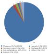
Figure 1
Figure 1
In the present literature survey, the duodenum was found to be the most common site of gangliocytic paraganglioma (90.2%, 229/254), followed by the respiratory system (2.4%, 6/254), low-level spinal cord (2.0%, 5/254), jejunum (1.2%, 3/254), esophagus (0.8%, 2/254), appendix (0.8%, 2/254), and others (2.8%, 7/254).
Figure 2
Figure 2
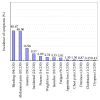
In the present literature survey, gastrointestinal bleeding was the most common symptom of gangliocytic paraganglioma (GP; 40.9%, 94/230), followed by abdominal pain (40.0%, 92/230), anemia (17.0%, 39/230), incidental findings (9.6%, 22/230), nausea (6.1%, 14/230), weight loss (4.8%, 11/230), and jaundice (4.4%, 10/230). It has been largely accepted that gastrointestinal bleeding and abdominal pain commonly occur in patients with GP; however, our survey revealed that obstructive jaundice is less common in patients with GP, although GP commonly occurs in the second part of the duodenum.
Figure 3
Figure 3
(a) Photomicrograph showing a low-power field of the gangliocytic paraganglioma (GP) site with a dense proliferation of epithelioid cells. Nested and compactly arranged epithelioid cells comprise the majority of the tumor (HE staining; magnification, ×100; the scale bar represents 300 μm). (b) Photomicrograph showing a high-power field of the GP site. The epithelioid cells have round to oval-shaped nuclei, inconspicuous nucleoli, and eosinophilic cytoplasm. Spindle-shaped cells surround the nests of epithelioid cells and are aligned in a single layer (HE staining; magnification, ×400; the scale bar represents 100 μm). (c) Photomicrograph showing a low-power field of the GP site with sporadic proliferation of epithelioid cells. A chaotic arrangement of epithelioid cells and a predominance of stromal cells are seen (HE staining; magnification, ×100; the scale bar represents 300 μm). (d) The epithelioid cells show a random arrangement, and spindle cells in the stroma are arranged in an irregular pattern (HE staining; magnification, ×400; the scale bar represents 100 μm).

References

    1. Hallet J., Law C. H. L., Cukier M., Saskin R., Liu N., Singh S. Exploring the rising incidence of neuroendocrine tumors: a population-based analysis of epidemiology, metastatic presentation, and outcomes. Cancer. 2015;121(4):589–597. doi: 10.1002/cncr.29099. - DOI - PubMed
    1. Öberg K., Castellano D. Current knowledge on diagnosis and staging of neuroendocrine tumors. Cancer and Metastasis Reviews. 2011;30, supplement 1(1):3–7. doi: 10.1007/s10555-011-9292-1. - DOI - PubMed
    1. Öberg K. E. Gastrointestinal neuroendocrine tumors. Annals of Oncology. 2010;21(7):vii72–vii80. doi: 10.1093/annonc/mdq290. - DOI - PubMed
    1. Okubo Y., Motohashi O., Nakayama N., et al. The clinicopathological significance of angiogenesis in hindgut neuroendocrine tumors obtained via an endoscopic procedure. Diagnostic Pathology. 2016;11, article 128 doi: 10.1186/s13000-016-0580-5. - DOI - PMC - PubMed
    1. Pape U.-F., Jann H., Müller-Nordhorn J., et al. Prognostic relevance of a novel TNM classification system for upper gastroenteropancreatic neuroendocrine tumors. Cancer. 2008;113(2):256–265. doi: 10.1002/cncr.23549. - DOI - PubMed

LinkOut - more resources