. 2016 Nov-Dec;10(11-12):E401-E403.
doi: 10.5489/cuaj.3402. Epub 2016 Nov 10.

Spontaneous uretero-sigmoid fistula secondary to calculus

Affiliations

Spontaneous uretero-sigmoid fistula secondary to calculus

Ines Marzouk et al. Can Urol Assoc J. 2016 Nov-Dec.

Abstract

A 25-year-old man was referred to the urology department after a subacute history of left back pain, burning micturition associated with pneumaturia and fecaluria. Ultrasonography was performed showing hydronephrosis, and plain film radiography demonstrated a long vertical left pelvic calculi. Uro-computed tomography (CT) combined with a water enema CT showed a 10 cm long calculus with the cranial extremity fistulating the sigmoidal wall. Surgical treatment included left nephroureterectomy and sigmoidectomy with a colorectal anastomosis. Postoperative course was uneventful.


Figures
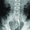
Fig. 1
Fig. 1
Plain film radiography showing a long stick-shaped vertical calcification in the left pelvic area (white arrow). Notice the other calcified opacities upper in the left kidney (black arrow).
Fig. 2
Fig. 2
Uro-computed tomography (CT) scans with water enema; (A) CT volume rendering scan at the delayed phase: non-functioning left kidney with multiple caliceal calculi; (B) multiplanar reformat CT scan showing the calculus presenting its cranial extremity in the sigmoidal lumen (arrow); (C) CT curved planar reformat in the axis of the left ureter showing a gas bubble (arrow) in the dilated ureter.
Fig. 3
Fig. 3
Intraoperative view after nephrectomy and before sigmoidectomy. Colonic fistula is well-identified with the calculus.

References

    1. Lee WK, Chang SD, Roche CJ, et al. Spontaneous ureterocolic fistula secondary to calculous pyohydroureter-onephrosis. Br J Radiol. 2005;78:954–5. doi: 10.1259/bjr/53711174. - DOI - PubMed
    1. Earlam MSS. Migration of a ureteric calculus to the bladder via a ureterocolic and a vesicocolic fistula. Aust N Z J Surg. 1977;47:519–20. doi: 10.1111/j.1445-2197.1977.tb04339.x. - DOI - PubMed
    1. Pai A, Coralic J, Park JJ, et al. Spontaneous ureterocolic fistula secondary to diverticulitis: Report of a case and review of literature. Int J Colorectal Dis. 2013;29:417–8. doi: 10.1007/s00384-013-1813-7. - DOI - PubMed
    1. Wc C, Sr P, Rw G. Spontaneous ureterocolic fistula: A rare complication of colonic diverticular disease. Am Surg. 1994;60:832–5. - PubMed
    1. Conde Santos G, Gri-ó Garreta J, Bielsa Gali O, et al. Ureterocolonic fistula in non-functioning ureter. Arch Esp Urol. 2001;54:1126–9. - PubMed

LinkOut - more resources