

In vitro effect of direct current electrical stimulation on rat mesenchymal stem cells
- PMID: 28097053
- PMCID: PMC5237370
- DOI: 10.7717/peerj.2821
In vitro effect of direct current electrical stimulation on rat mesenchymal stem cells
Abstract
Background: Electrical stimulation (ES) has been successfully used to treat bone defects clinically. Recently, both cellular and molecular approaches have demonstrated that ES can change cell behavior such as migration, proliferation and differentiation.
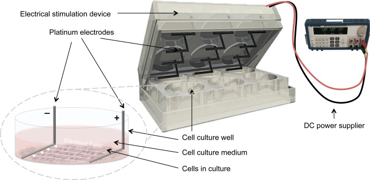
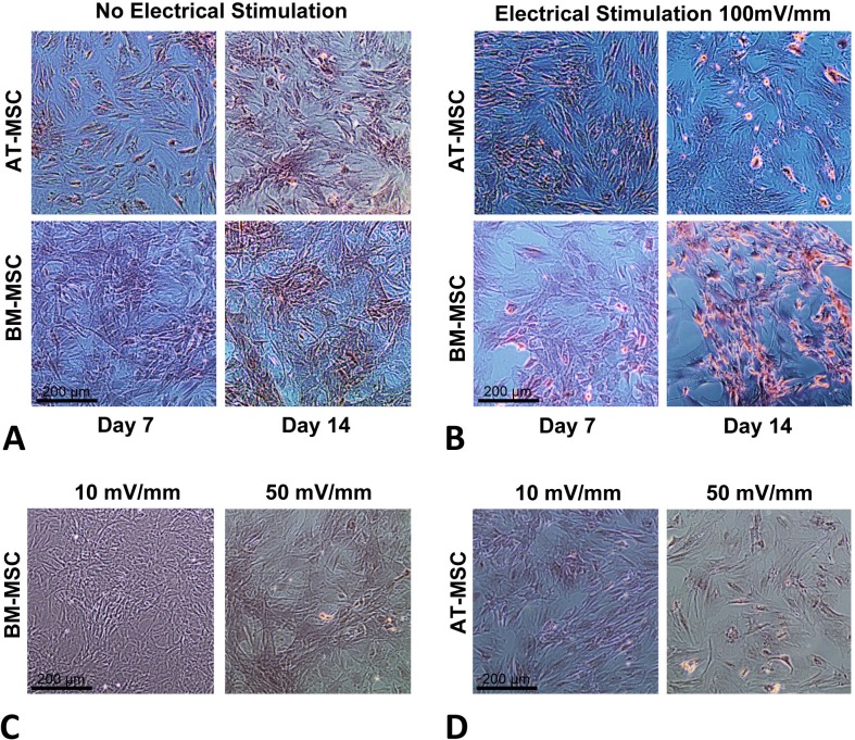
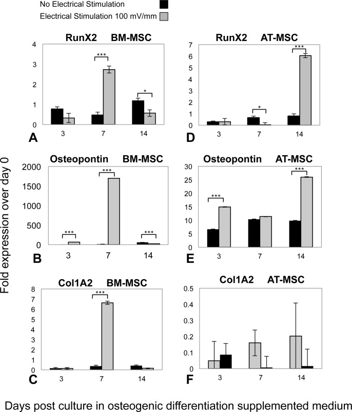
Methods: In the present study we exposed rat bone marrow- (BM-) and adipose tissue- (AT-) derived mesenchymal stem cells (MSCs) to direct current electrical stimulation (DC ES) and assessed temporal changes in osteogenic differentiation. We applied 100 mV/mm of DC ES for 1 h per day for three, seven and 14 days to cells cultivated in osteogenic differentiation medium and assessed viability and calcium deposition at the different time points. In addition, expression of osteogenic genes, Runx2, Osteopontin, and Col1A2 was assessed in BM- and AT-derived MSCs at the different time points.
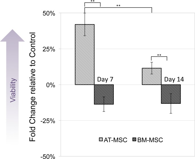
Results: Results showed that ES changed osteogenic gene expression patterns in both BM- and AT-MSCs, and these changes differed between the two groups. In BM-MSCs, ES caused a significant increase in mRNA levels of Runx2, Osteopontin and Col1A2 at day 7, while in AT-MSCs, the increase in Runx2 and Osteopontin expression were observed after 14 days of ES.
Discussion: This study shows that rat bone marrow- and adipose tissue-derived stem cells react differently to electrical stimuli, an observation that could be important for application of electrical stimulation in tissue engineering.
Keywords: Adipose tissue-derived mesenchymal stem cells; Bone marrow-derived mesenchymal stem cells; Bone tissue engineering; Direct current electrical stimulation.
Conflict of interest statement
The authors declare there are no competing interests.
Figures
References
-
- Balint R, Cassidy NJ, Hidalgo-Bastida LA, Cartmell S. Electrical stimulation enhanced mesenchymal stem cell gene expression for orthopaedic tissue repair. Journal of Biomaterials and Tissue Engineering. 2013;3:212–221. doi: 10.1166/jbt.2013.1081. - DOI
-
- Bodamyali T, Bhatt B, Hughes FJ, Winrow VR, Kanczler JM, Simon B, Abbott J, Blake DR, Stevens CR. Pulsed electromagnetic fields simultaneously induce osteogenesis and upregulate transcription of bone morphogenetic proteins 2 and 4 in rat osteoblasts in vitro. Biochemical and Biophysical Research Communications. 1998;250:458–461. doi: 10.1006/bbrc.1998.9243. - DOI - PubMed
LinkOut - more resources
Full Text Sources
Other Literature Sources
Research Materials
Miscellaneous
