. 2016 Dec 14;7(1):74-87.
doi: 10.1002/2211-5463.12157. eCollection 2017 Jan.

A comparative study of the degradation of yeast cyclins Cln1 and Cln2

Affiliations

A comparative study of the degradation of yeast cyclins Cln1 and Cln2

Inma Quilis et al. FEBS Open Bio. .

Abstract

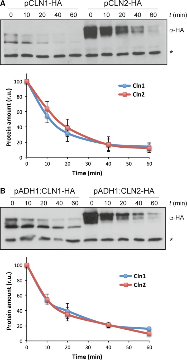
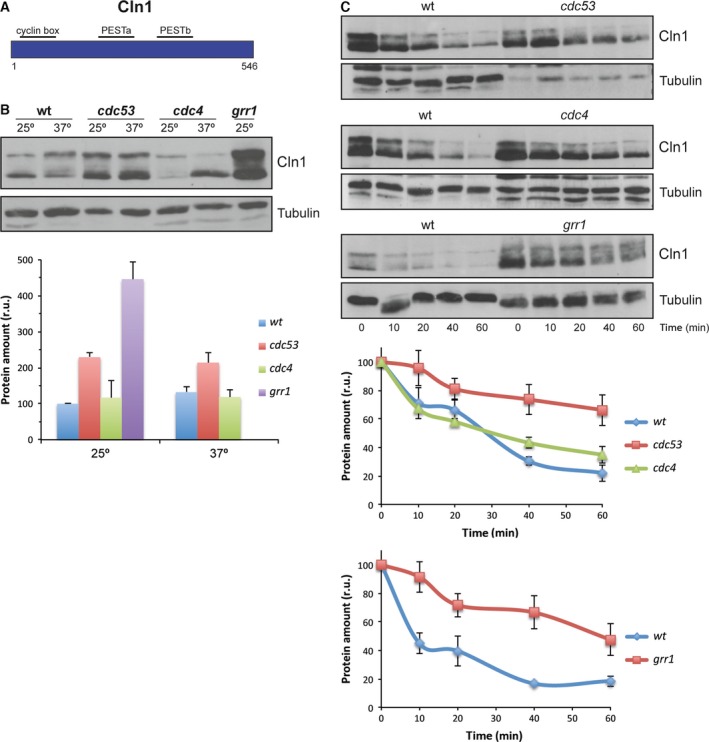
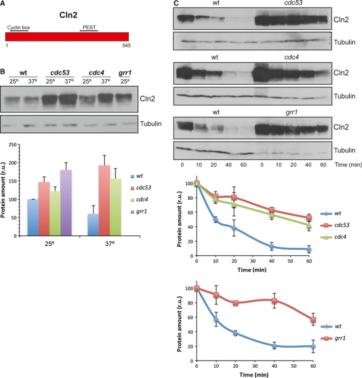
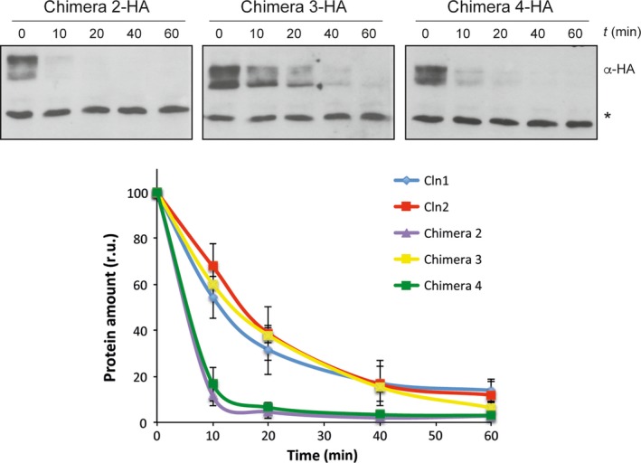
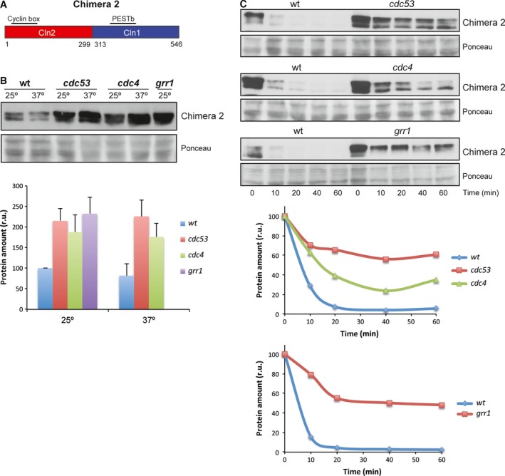
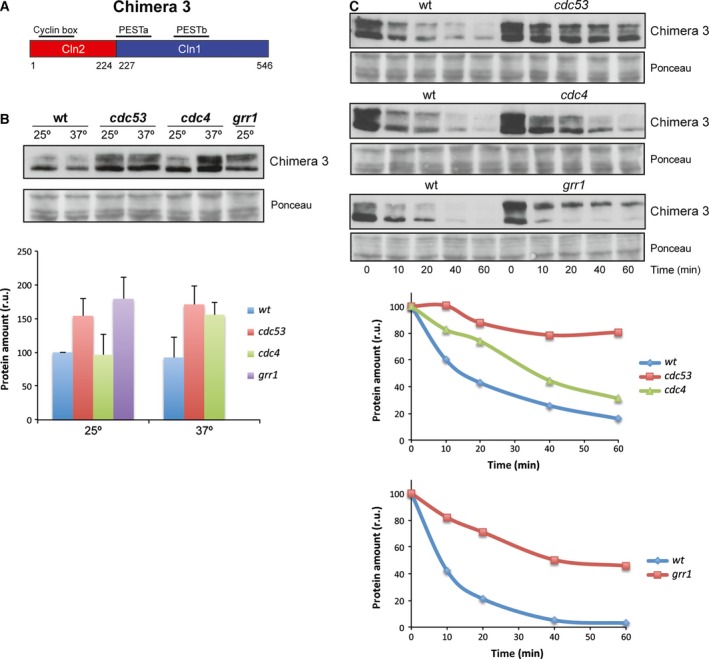
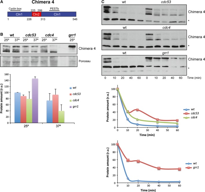
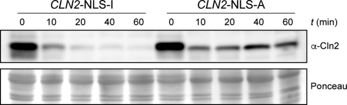
The yeast cyclins Cln1 and Cln2 are very similar in both sequence and function, but some differences in their functionality and localization have been recently described. The control of Cln1 and Cln2 cellular levels is crucial for proper cell cycle initiation. In this work, we analyzed the degradation patterns of Cln1 and Cln2 in order to further investigate the possible differences between them. Both cyclins show the same half-life but, while Cln2 degradation depends on ubiquitin ligases SCFGrr1 and SCFCdc4, Cln1 is affected only by SCFGrr1. Degradation analysis of chimeric cyclins, constructed by combining fragments from Cln1 and Cln2, identifies the N-terminal sequence of the proteins as responsible of the cyclin degradation pattern. In particular, the N-terminal region of Cln2 is required to mediate degradation by SCFCdc4. This region is involved in nuclear import of Cln1 and Cln2, which suggests that differences in degradation may be due to differences in localization. Moreover, a comparison of the cyclins that differ only in the presence of the Cln2 nuclear export signal indicates a greater instability of exported cyclins, thus reinforcing the idea that cyclin stability is influenced by their localization.

Keywords: Cln1; Cln2; SCF ubiquitin ligase; Saccharomyces cerevisiae; cell cycle; cyclin.


Figures

Figure 1
Figure 1
Protein stability of cyclins Cln1 and Cln2. Exponentially growing cells of the wild‐type strain transformed with a centromeric plasmid expressing either a HA‐epitope tagged Cln1 or Cln2 protein at endogenous level (A) or overexpressed from the ADH1 promoter (B) were incubated in the presence of 50 μg·mL−1 cycloheximide. Cln1 and Cln2 protein levels were analyzed at the indicated time after the addition of cycloheximide by western blot. A nonspecific band labeled with an asterisk that cross‐react with the antibody is shown as loading control. Graph represents the relative amount of Cln1 and Cln2 proteins related to the nonspecific band.
Figure 2
Figure 2
Analysis of Cln1 degradation in SCF ubiquitin‐ligase mutant strains. (A) Scheme of Cln2 cyclin. (B) Cells of the wild‐type (W303), cdc53, cdc4 and grr1 (JCY1539) strains transformed with a centromeric plasmid expressing a HA‐epitope tagged Cln1 protein at endogenous level were grown at 25° and transferred for 3 h at 37° in the case of cdc53 and cdc4 strains. Cln1 protein level was analyzed by western blot. Tubulin is shown as loading control. Graph represents the relative amount of Cln1 protein. (C) Same cells than in B were incubated in the presence of 50 μg·mL−1 cycloheximide. Cln1 protein level was analyzed at the indicated time after the addition of cycloheximide by western blot. Tubulin is shown as loading control. Graph represents the relative amount of Cln1 protein.
Figure 3
Figure 3
Analysis of Cln2 degradation in SCF ubiquitin‐ligase mutant strains. (A) Scheme of Cln2 cyclin. (B, C) Protein level and degradation of Cln2 cyclin were analyzed as described in Fig. 2.
Figure 4
Figure 4
Protein stability of chimeric cyclins. Protein stability of chimeric cyclins was analyzed as described in Fig. 1.
Figure 5
Figure 5
Analysis of Chimera 2 degradation in SCF ubiquitin‐ligase mutant strains. (A) Scheme of Chimera 2 cyclin. (B, C) Protein level and degradation of Chimera 2 cyclin were analyzed as described in Fig. 2. Ponceau staining is shown as loading control.
Figure 6
Figure 6
Analysis of Chimera 3 degradation in SCF ubiquitin‐ligase mutant strains. (A) Scheme of Chimera 3 cyclin. (B, C) Protein level and degradation of Chimera 3 cyclin were analyzed as described in Fig. 2. Ponceau staining is shown as loading control.
Figure 7
Figure 7
Analysis of Chimera 4 degradation in SCF ubiquitin‐ligase mutant strains. (A) Scheme of Chimera 4 cyclin. (B, C) Protein level and degradation of Chimera 4 cyclin were analyzed as described in Fig. 2. Ponceau staining or a nonspecific band labeled with an asterisk that cross‐react with the antibody is shown as loading control.
Figure 8
Figure 8
Protein stability of Cln2 protein with altered localization. Protein level of Cln2 cyclin fused to two copies of either active or inactive SV40 NLS was analyzed at the indicated time after the addition of cycloheximide by western blot. Ponceau staining is shown as loading control.

Similar articles

Cited by

References

    1. Andrews B and Measday V (1998) The cyclin family of budding yeast: abundant use of a good idea. Trends Genet 14, 66–72. - PubMed
    1. Mendenhall MD and Hodge AE (1998) Regulation of Cdc28 cyclin‐dependent protein kinase activity during the cell cycle of the yeast Saccharomyces cerevisiae . Microbiol Mol Biol Rev 62, 1191–1243. - PMC - PubMed
    1. Breeden LL (2003) Periodic transcription: a cycle within a cycle. Curr Biol 13, R31–R38. - PubMed
    1. McInerny CJ (2011) Cell cycle regulated gene expression in yeasts. Adv Genet 73, 51–85. - PubMed
    1. Simon I, Barnett J, Hannett N, Harbison CT, Rinaldi NJ, Volkert TL, Wyrick JJ, Zeitlinger J, Gifford DK, Jaakkola TS et al (2001) Serial regulation of transcriptional regulators in the yeast cell cycle. Cell 106, 697–708. - PubMed

LinkOut - more resources