

Establishment of a Novel Simplified Surgical Model of Acute Liver Failure in the Cynomolgus Monkey
- PMID: 28097130
- PMCID: PMC5209601
- DOI: 10.1155/2016/3518989
Establishment of a Novel Simplified Surgical Model of Acute Liver Failure in the Cynomolgus Monkey
Abstract
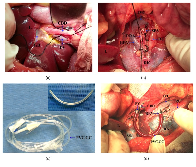
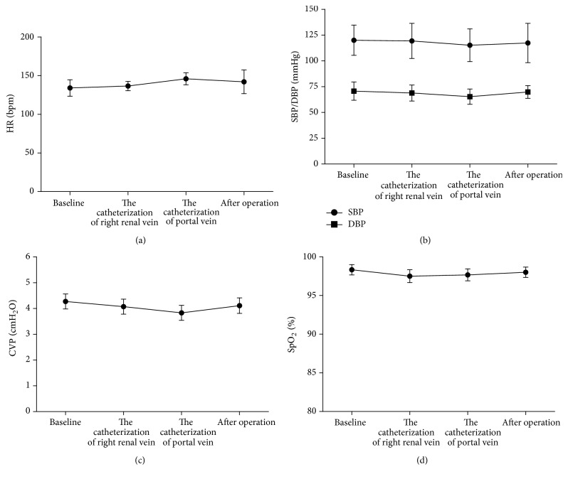
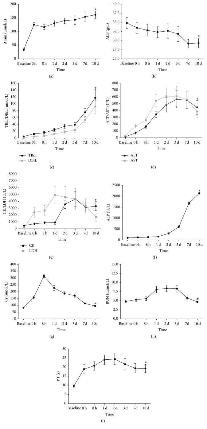
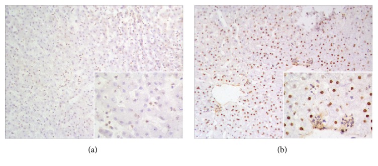
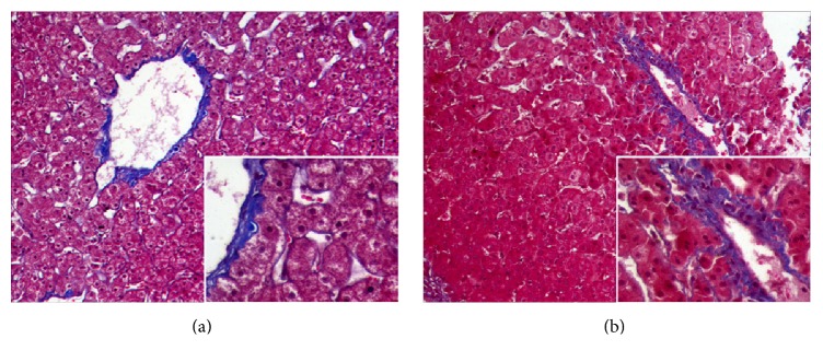
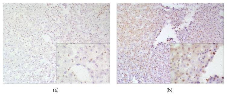
Models using large animals that are suitable for studying artificial liver support system (ALSS) are urgently needed. Presently available acute liver failure (ALF) models mainly involve pigs or dogs. Establishment of current surgical ALF models (hepatectomy/devascularization) requires either very good surgical skills or multistep processes-even multiple stages of surgery. Therefore, it is necessary to develop a simplified surgical method. Here we report a novel simplified surgical ALF model using cynomolgus monkeys. Six monkeys underwent portal-right renal venous shunt combined with common bile duct ligation and transection (PRRS + CBDLT). Postoperatively, the monkeys had progressively increased listlessness, loss of appetite, and obvious jaundice. Blood biochemistry levels (Amm, ALT, AST, TBiL, DBiL, ALP, LDH, CK, and Cr) and prothrombin time (PT) were significantly increased (all P < 0.01) and albumin (ALB) was markedly reduced (P < 0.01) compared with baseline values. Histological examination of liver specimens on postoperative day 10 revealed cholestasis and inflammation. PRRS + CBDLT produced ALF that closely correlated with clinical situations. Compared with other surgical or drug ALF models, ours was simplified and animals were hemodynamically stable. This model could provide a good platform for further research on ALSS, especially regarding their detoxification functions.
Conflict of interest statement
All authors of this article declare that they have no conflict of interests.
Figures

Similar articles
-
Novel D-galactosamine-induced cynomolgus monkey model of acute liver failure.World J Gastroenterol. 2017 Nov 14;23(42):7572-7583. doi: 10.3748/wjg.v23.i42.7572. World J Gastroenterol. 2017. PMID: 29204057 Free PMC article.
-
Porcine acute liver failure model established by two-phase surgery and treated with hollow fiber bioartificial liver support system.World J Gastroenterol. 2005 Sep 21;11(35):5468-74. doi: 10.3748/wjg.v11.i35.5468. World J Gastroenterol. 2005. PMID: 16222738 Free PMC article.
-
Experimental research on TECA-I bioartificial liver support system to treat canines with acute liver failure.World J Gastroenterol. 2001 Oct;7(5):706-9. doi: 10.3748/wjg.v7.i5.706. World J Gastroenterol. 2001. PMID: 11819859 Free PMC article.
-
A novel stable reproducible model of hepatic failure in canines.J Surg Res. 2000 Dec;94(2):167-71. doi: 10.1006/jsre.2000.5997. J Surg Res. 2000. PMID: 11104657
-
Acute liver failure: a critical appraisal of available animal models.Metab Brain Dis. 2005 Dec;20(4):409-23. doi: 10.1007/s11011-005-7927-z. Metab Brain Dis. 2005. PMID: 16382351 Review.
Cited by
-
Novel D-galactosamine-induced cynomolgus monkey model of acute liver failure.World J Gastroenterol. 2017 Nov 14;23(42):7572-7583. doi: 10.3748/wjg.v23.i42.7572. World J Gastroenterol. 2017. PMID: 29204057 Free PMC article.
-
Stem Cell-Based Strategies: The Future Direction of Bioartificial Liver Development.Stem Cell Rev Rep. 2024 Apr;20(3):601-616. doi: 10.1007/s12015-023-10672-5. Epub 2024 Jan 3. Stem Cell Rev Rep. 2024. PMID: 38170319 Review.
-
Hepatobiliary organoids differentiated from hiPSCs relieve cholestasis-induced liver fibrosis in nonhuman primates.Int J Biol Sci. 2024 Jan 21;20(4):1160-1179. doi: 10.7150/ijbs.90441. eCollection 2024. Int J Biol Sci. 2024. PMID: 38385067 Free PMC article.
-
A novel, simplified, and reproducible porcine model of acute ischemic liver failure with portal vein preservation.Exp Anim. 2022 Feb 9;71(1):60-70. doi: 10.1538/expanim.21-0076. Epub 2021 Sep 8. Exp Anim. 2022. PMID: 34497163 Free PMC article.
References
-
- Saliba F., Samuel D. Artificial liver support: a real step forward. Minerva Medica. 2015;106(1):35–43. - PubMed
MeSH terms
LinkOut - more resources
Full Text Sources
Other Literature Sources
Research Materials
Miscellaneous
