

Fully automated quantitative cephalometry using convolutional neural networks
- PMID: 28097213
- PMCID: PMC5220585
- DOI: 10.1117/1.JMI.4.1.014501
Fully automated quantitative cephalometry using convolutional neural networks
Abstract
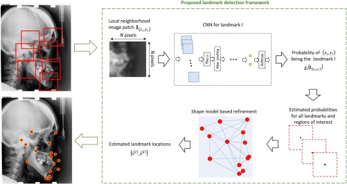
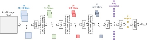
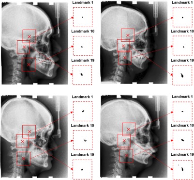
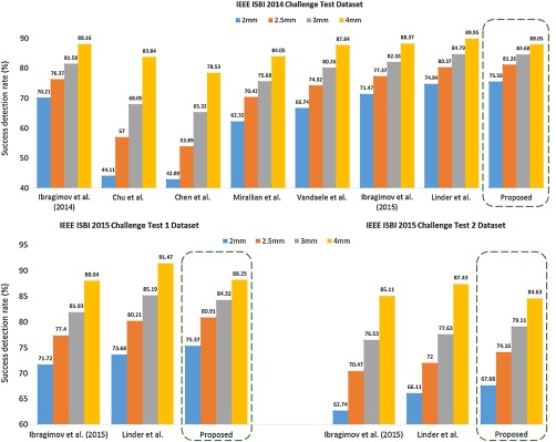
Quantitative cephalometry plays an essential role in clinical diagnosis, treatment, and surgery. Development of fully automated techniques for these procedures is important to enable consistently accurate computerized analyses. We study the application of deep convolutional neural networks (CNNs) for fully automated quantitative cephalometry for the first time. The proposed framework utilizes CNNs for detection of landmarks that describe the anatomy of the depicted patient and yield quantitative estimation of pathologies in the jaws and skull base regions. We use a publicly available cephalometric x-ray image dataset to train CNNs for recognition of landmark appearance patterns. CNNs are trained to output probabilistic estimations of different landmark locations, which are combined using a shape-based model. We evaluate the overall framework on the test set and compare with other proposed techniques. We use the estimated landmark locations to assess anatomically relevant measurements and classify them into different anatomical types. Overall, our results demonstrate high anatomical landmark detection accuracy ([Formula: see text] to 2% higher success detection rate for a 2-mm range compared with the top benchmarks in the literature) and high anatomical type classification accuracy ([Formula: see text] average classification accuracy for test set). We demonstrate that CNNs, which merely input raw image patches, are promising for accurate quantitative cephalometry.
Keywords: artificial neural networks; feed-forward neural networks; image recognition; machine vision; predictive models; statistical learning; supervised learning; x-ray applications.
Figures
References
-
- Kafieh R., et al. , “Discrimination of bony structures in cephalograms for automatic landmark detection,” in Advances in Computer Science and Engineering, Sarbazi-Azad H., et al., Eds., pp. 609–620, Springer, Berlin Heidelberg: (2008).
-
- Kaur A., Singh C., “Automatic cephalometric landmark detection using Zernike moments and template matching,” Signal Image Video Process. 9(1), 117–132 (2015).10.1007/s11760-013-0432-7 - DOI
Grants and funding
LinkOut - more resources
Full Text Sources
Other Literature Sources
