Comparative Study
. 2017 Jan 12;2(1):e90139.
doi: 10.1172/jci.insight.90139.

Oxidized CaMKII promotes asthma through the activation of mast cells

Affiliations
Comparative Study

Oxidized CaMKII promotes asthma through the activation of mast cells

Jingjing Qu et al. JCI Insight. .

Abstract

Oxidation of calmodulin-dependent protein kinase II (ox-CaMKII) by ROS has been associated with asthma. However, the contribution of ox-CaMKII to the development of asthma remains to be fully characterized. Here, we tested the effect of ox-CaMKII on IgE-mediated mast cell activation in an allergen-induced mouse model of asthma using oxidant-resistant CaMKII MMVVδ knockin (MMVVδ) mice. Compared with WT mice, the allergen-challenged MMVVδ mice displayed less airway hyperresponsiveness (AHR) and inflammation. These MMVVδ mice exhibited reduced levels of ROS and diminished recruitment of mast cells to the lungs. OVA-activated bone marrow-derived mast cells (BMMCs) from MMVVδ mice showed a significant inhibition of ROS and ox-CaMKII expression. ROS generation was dependent on intracellular Ca2+ concentration in BMMCs. Importantly, OVA-activated MMVVδ BMMCs had suppressed degranulation, histamine release, leukotriene C4, and IL-13 expression. Adoptive transfer of WT, but not MMVVδ, BMMCs, reversed the alleviated AHR and inflammation in allergen-challenged MMVVδ mice. The CaMKII inhibitor KN-93 significantly suppressed IgE-mediated mast cell activation and asthma. These studies support a critical but previously unrecognized role of ox-CaMKII in mast cells that promotes asthma and suggest that therapies to reduce ox-CaMKII may be a novel approach for asthma.


Conflict of interest statement

M.E. Anderson is a cofounder of Allosteros Therapeutics, a biotech company aiming to develop CaMKII-based therapies.

Figures

Figure 1
Figure 1. Ox-CaMKII regulates cockroach allergen-induced airway hyperresponsiveness and inflammation.
(A) Protocol for cockroach allergen-induced (CRE-induced) mouse model of asthma. (B) Paraffin tissue sections of lung were stained with H&E (top; scale bar: 100 μm) and periodic acid–Schiff (PAS, bottom; scale bar: 50 μm). (C and D) Lung resistance (C) and compliance (D) in response to increasing concentrations of methacholine using the forced oscillation technique (FlexiVent, SCIREQ) from CRE-challenged WT (n = 6) and MMVVδ (n = 6) mice. (E and F) Bronchoalveolar lavage (BAL) total (E) and differential cell counts (F) of PBS- (n = 7) and CRE-challenged WT (n = 11) and MMVVδ (n = 11) mice. (G) Serum levels of cockroach allergen-specific IgE and IgG1. (H) Levels of cytokines in BAL. Data represent mean ± SEM; comparisons were made using 2-tailed Student’s t test between CRE-treated WT vs. MMVVδ. *P < 0.05, **P < 0.01, ***P < 0.001.
Figure 2
Figure 2. ROS-resistant CaMKII MMVVδ mice prevent ROS production and oxidative activity of CaMKII.
(A) Representative images of dihydroethidium-stained (DHE-stained) airways of PBS- or CRE-challenged WT and MMVVδ mice. Scale bar: 100 μm (B) Representative immunoblot of ox-CaMKII and total CaMKII (Tot-CaMKII) in the homogenized lung tissues of PBS- or CRE-challenged WT and MMVVδ mice. Data represent mean ± SEM, 3 mice per group. Comparisons were made using 2-tailed Student’s t test between CRE-treated WT vs. MMVV. **P < 0.01, ***P < 0.001.
Figure 3
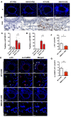
Figure 3. Reduced mast cell numbers in lung tissues of CRE-treated MMVVδ mice.
(A and B) Representative images of tryptase immunofluorescence (scale bar: 100 μm) and DAB staining (scale bar: 50 μm) in lung tissue sections of PBS- or CRE-challenged WT and MMVVδ mice, 3 mice per group. (C and D) Quantitative data for immunofluorescence (C) and DAB staining (D). Mean ± SEM, n = 5–9 high power field view (HFV) per group. (E) Representative images of coimmunofluorescence staining with c-Kit and ox-CaMKII in the lung tissues of CRE-challenged WT and MMVVδ mice, 3 mice per group. Scale bar: 100 μm (first and third row); 20 μm (second and fourth row). Quantitative data for c-Kit+ cells (F) and ox-CaMKII+c-Kit+ cells (G). Data represent mean ± SEM, n = 7–9 HFV per group. Comparisons were made using 2-tailed Student’s t test between CRE-treated WT vs. MMVVδ. *P < 0.05, **P < 0.01.
Figure 4
Figure 4. Reduced ROS levels in mast cells of ROS-resistant CaMKII mice.
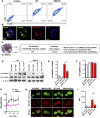
(A) Representation of BMMC FACS analysis for the surface markers c-Kit and FcεRI. (B) Representation of images of the colocalization of CaMKII (green) and mitochondria (red) in BMMCs using confocal microscopy. Representative of 2 independent experiments. Scale bar: 10 μm. (C) Experimental setup for IgE-mediated mast cell activation and expression of CaMKII and ROS. BMMCs from WT and MMVVδ mice were sensitized with 1 μg/ml anti-OVA IgE (E-C1) for 16 hours and then stimulated with 10 μg/ml OVA for 30 minutes. (D) Representative immunoblot of ox-CaMKII and total CaMKII (Tot-CaMKII) in OVA-sensitized and challenged BMMCs derived from WT and MMVVδ mice. Various dosages of H2O2 were used as possible controls. One representative immunoblot of three is shown. (E and F) Quantitative data for ox-CaMKII (E) and Tot-CaMKII (F) expression using densitometry from 3 independent Western blots. Mean ± SEM. (G) Levels of intracellular ROS in OVA-sensitized and challenged BMMCs by flow cytometry with CM-H2DCFDA. Mean ± SEM, a single time point from 3 independent experiments. (H) Representative MitoTracker or MitoSOX staining of these OVA-sensitized and challenged WT and MMVVδ BMMCs from 3 independent experiments. Scale bar: 5 μm. (I) Quantitative data for MitoSOX expression in H, as determined by mean fluorescent intensity (MFI). Data represent mean ± SEM, 7–14 high power field view (HFV) per group. Comparisons were made using 2-tailed Student’s t test between OVA-treated WT vs. MMVV. *P < 0.05.
Figure 5
Figure 5. ROS production is dependent on intracellular Ca2+.
(A) Representative Fluo-4 fluorescence heatmap images of anti-OVA IgE-sensitized BMMCs showing changes in [Ca2+]i induced by OVA. Representative of 3 independent experiments. Scale bar: 20 μm. (B) Representative mean fluorescent intensity (MFI) average traces for sensitized and challenged BMMCs from WT and MMVV BMMCs. CIB, Ca2+ imaging buffer. (C) Quantification of total calcium response from sensitized and challenged cells (>150 cells counted per condition) by calculating the AUC. (D and E) Levels of intracellular ROS in OVA-sensitized and challenged WT (D) or MMVVδ (E) BMMCs in the presence of IP3 receptor antagonist 2-APB by flow cytometry with CM-H2DCFDA. Data represent mean ± SEM, a single time point from 3 independent experiments. Comparisons were made using 2-tailed Student’s t test between OVA-treated WT vs. MMVV group (C) or 2-APB–treated vs. nontreated OVA-activated WT (D) and MMVVδ BMMCs (E). *P < 0.05, **P < 0.01.
Figure 6
Figure 6. Oxidized CaMKII can modulate allergen-induced mast cell activation.
(AD) Levels of β-hexosaminidase (A), histamine (B), LTC4 (C), and IL-13 (D) in sensitized and challenged WT and MMVVδ BMMCs detected by ELISA. Mean ± SEM, n = 4 per group. (E) Experimental setup for IgE-mediated passive cutaneous anaphylaxis (PCA). Mice were injected intradermally with E-C1. After 48 hours, OVA was administered i.v. together with Evans blue dye for 30 minutes, followed by the quantification of the extravasation of Evans blue leakage into the skin. (F) Representative images of Evans blue–stained extravasation into skin 30 minutes after i.v. injection of OVA from WT and MMVVδ mice. For quantification of the extravasation of Evans blue leakage into the skin. Absorbance was measured at 620 nm, and data are expressed as Evans blue in ng/mg tissue. Data represent mean ± SEM, n = 4 per group. Comparisons were made using 2-tailed Student’s t test between OVA-treated WT vs. MMVV group. *P < 0.05, **P < 0.01.
Figure 7
Figure 7. Adoptive transfer of WT mast cells reverses the protective effect of CaMKII MMVVδ in a mouse model of asthma.
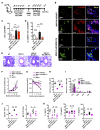
(A) Protocol for adaptive transfer of BMMCs in the cockroach allergen-induced mouse model of asthma. (B) Colocalization of c-Kit (green) and ox-CaMKII (red) in the lung sections of MMVVδ mice with adoptively transferred WT and MMVVδ BMMCs. Scale bar: 100 μm (first and third row); 20 μm (second and fourth row). (C and D) Numbers of c-Kit–positive cells (C) and cells positive for both c-Kit and ox-CaMKII (D) from WT and MMVVδ mice with adoptive transfer of WT and MMVVδ BMMCs. Mean ± SEM, n = 8 high power field view (HFV) per group. (E) Representation of H&E-stained paraffin lung tissue sections from WT and MMVVδ mice adoptively transferred with WT or MMVVδ BMMCs in a CRE-induced mouse model of asthma (6 mice per group). Scale bar: 100 μm. (F and G) Systemic airway resistance (F) and compliance (G) in response to increasing concentrations of methacholine using the forced oscillation technique (FlexiVent, SCIREQ). Data represent mean ± SEM, 6 mice per group. (H and I) Bronchoalveolar lavage (BAL) total (H) and differential (I) cell counts of CRE-challenged WT and MMVVδ mice. Mean ± SEM, 7 mice per group. (J) Serum levels of cockroach allergen-specific IgE and IgG1. Mean ± SEM, 7 mice per group. (K) Levels of IL-4 (n = 7), IL-5 (n = 12), and IL-13 (n = 12) in BALs. Data represent mean ± SEM; comparisons were made using 2-tailed Student’s t test of CRE-treated MMVVδ vs. MMVVδ with adoptively transferred WT BMMCs and MMVVδ with adoptively transferred WT vs. MMVVδ BMMCs. *P < 0.05, **P < 0.01, ***P < 0.001.
Figure 8
Figure 8. CaMKII inhibitor suppresses OVA-induced mast cell activation and PCA in vivo.
BMMCs or human mast cells, HMC-1 cells, were sensitized with 1 μg/ml anti-OVA IgE (E-C1) in the presence or absence of different doses of KN-93 (1–20 μM) for 16 hours and then stimulated with PBS or 10 μg/ml OVA for 30 minutes. (AD) Mast cell activation was assessed by measuring β-hexosaminidase (n = 3) in BMMCs (A) and HMC-1 cells (C) and IL-13 levels (n = 6) in BMMCs (B) and HMC-1 cells (D). (E) Representative images of Evans blue–stained extravasation into skin. Mice were injected intradermally with E-C1 with or without KN-93. After 24 hours, OVA was administered i.v. together with Evans blue dye for 30 minutes, followed by the quantification of the extravasation of Evans blue leakage into the skin. Data represent mean ± SEM, 3 mice per group. Comparisons were made using 2-tailed Student’s t test between OVA-treated mast cells vs. mast cells pretreated with different dosages of KN-93 and then challenged with OVA (AD) or mice pretreated with or without KN-93 and then challenged with OVA (E). *P < 0.05, **P < 0.01.

Similar articles

Cited by

References

    1. Jarjour NN, Calhoun WJ. Enhanced production of oxygen radicals in asthma. J Lab Clin Med. 1994;123(1):131–136. - PubMed
    1. Abdala-Valencia H, et al. Nonhematopoietic NADPH oxidase regulation of lung eosinophilia and airway hyperresponsiveness in experimentally induced asthma. Am J Physiol Lung Cell Mol Physiol. 2007;292(5):L1111–L1125. doi: 10.1152/ajplung.00208.2006. - DOI - PMC - PubMed
    1. Bowler RP. Oxidative stress in the pathogenesis of asthma. Curr Allergy Asthma Rep. 2004;4(2):116–122. doi: 10.1007/s11882-004-0056-7. - DOI - PubMed
    1. Li YJ, Takizawa H, Kawada T. Role of oxidative stresses induced by diesel exhaust particles in airway inflammation, allergy and asthma: their potential as a target of chemoprevention. Inflamm Allergy Drug Targets. 2010;9(4):300–305. doi: 10.2174/187152810793358787. - DOI - PubMed
    1. Al-Harbi NO, et al. Airway oxidative stress causes vascular and hepatic inflammation via upregulation of IL-17A in a murine model of allergic asthma. Int Immunopharmacol. 2016;34:173–182. doi: 10.1016/j.intimp.2016.03.003. - DOI - PubMed

Publication types

MeSH terms