. 2017 Dec;5(1):3.
doi: 10.1186/s40635-016-0115-0. Epub 2017 Jan 17.

Deletion of Nlrp3 protects from inflammation-induced skeletal muscle atrophy

Affiliations

Deletion of Nlrp3 protects from inflammation-induced skeletal muscle atrophy

Nora Huang et al. Intensive Care Med Exp. 2017 Dec.

Abstract

Background: Critically ill patients develop atrophic muscle failure, which increases morbidity and mortality. Interleukin-1β (IL-1β) is activated early in sepsis. Whether IL-1β acts directly on muscle cells and whether its inhibition prevents atrophy is unknown. We aimed to investigate if IL-1β activation via the Nlrp3 inflammasome is involved in inflammation-induced atrophy.

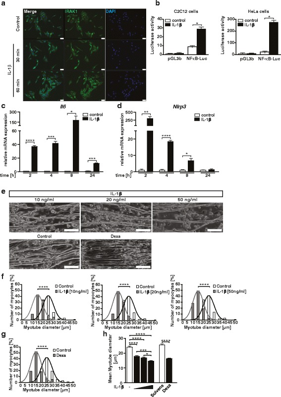
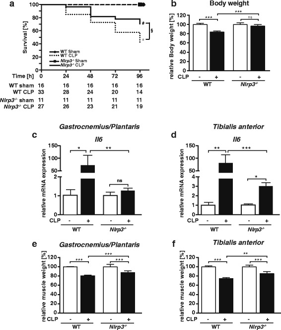
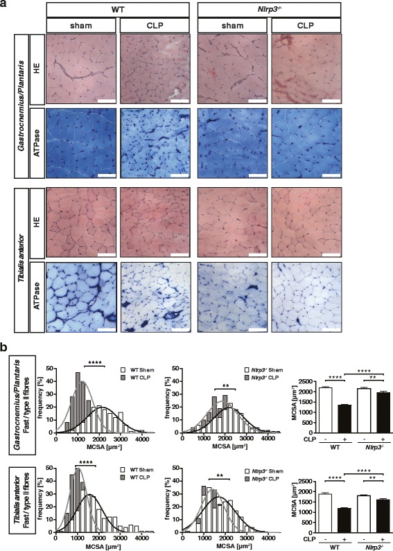
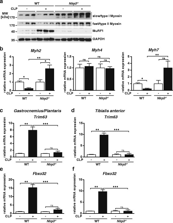
Methods: We performed an experimental study and prospective animal trial. The effect of IL-1β on differentiated C2C12 muscle cells was investigated by analyzing gene-and-protein expression, and atrophy response. Polymicrobial sepsis was induced by cecum ligation and puncture surgery in Nlrp3 knockout and wild type mice. Skeletal muscle morphology, gene and protein expression, and atrophy markers were used to analyze the atrophy response. Immunostaining and reporter-gene assays showed that IL-1β signaling is contained and active in myocytes.

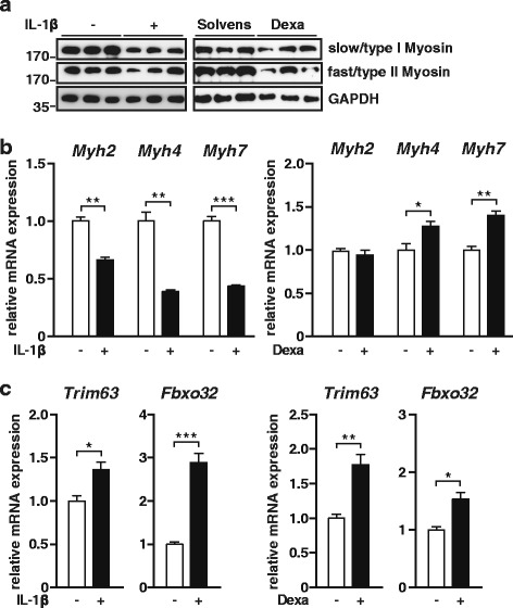
Results: Immunostaining and reporter gene assays showed that IL-1β signaling is contained and active in myocytes. IL-1β increased Il6 and atrogene gene expression resulting in myocyte atrophy. Nlrp3 knockout mice showed reduced IL-1β serum levels in sepsis. As determined by muscle morphology, organ weights, gene expression, and protein content, muscle atrophy was attenuated in septic Nlrp3 knockout mice, compared to septic wild-type mice 96 h after surgery.

Conclusions: IL-1β directly acts on myocytes to cause atrophy in sepsis. Inhibition of IL-1β activation by targeting Nlrp3 could be useful to prevent inflammation-induced muscle failure in critically ill patients.

Keywords: ICUAW; IL-1β; Muscle weakness; Sepsis.


Figures

Fig. 1
Fig. 1
The IL-1β signaling pathway is contained and active in C2C12 myocytes. a C2C12 muscle cells were treated with human recombinant IL-1β (10 ng/ml) or vehicle for 30 min and 1 h. Immunocytochemistry with anti-IRAK1 antibody shows cytoplasmic-to-nuclear translocation of IRAK1 in response to IL-1β after 30 min. Nuclei were stained in blue (DAPI). Scale bar = 50 μm. b Synthetic luciferase reporters with multimerized NF-κB sites (NF-κB-Luc) were transfected into C2C12 (b, left panel) and HeLa (b, right panel) cells, together with LacZ as transfection control. Cells were treated with recombinant IL-1β (10 ng/ml) for 24 h. n = 3. c, d C2C12 cells were differentiated for 8 days and treated with human recombinant IL-1β (10 ng/ml) for different time points as indicated. qRT-PCR analysis of Il6 and Nlrp3. mRNA expression was normalized to Gapdh. All data are reported as fold change ± SEM. e–h IL-1β increases Nlrp3 expression and induces atrophy in differentiated C2C12 myocytes in vitro. C2C12 cells were differentiated for 8 days and treated with human recombinant IL-1β (10, 20, and 50 ng/ml) for 72 h. Dexamethasone (10 μM/ml) treatment was used as atrophy control. e Representative light microscopy pictures. Scale bar = 250 μm. f, g Frequency distribution histograms of cell width of IL-1β (10, 20, and 50 ng/ml) and dexamethasone-treated myotubes, as indicated, compared to vehicle-treated myotubes, n = 100 cells per condition. h Mean myotube width. Data are presented as mean ± SEM. *p < 0.05, **p < 0.01, ***p < 0.001, ****p ≤ 0.0001
Fig. 2
Fig. 2
IL-1β treatment induces Trim63 (MuRF1) and Fbxo32 (atrogin 1) gene expression and reduces slow and fast myosin heavy chain (MyHC) in C2C12 myotubes. a C2C12 cells were differentiated for 8 days and treated with human recombinant IL-1β (10 ng/ml) for 72 h. Dexamethasone (10 μM/ml) treatment was used as atrophy control. Western blot analysis with anti-myosin heavy chain (MyHC) slow and anti-MyHC-fast antibody. n = 3. GAPDH was used as loading control. b C2C12 cells were differentiated for 8 days and treated with human recombinant IL-1β (10 ng/ml) for 24 h. Dexamethasone (10 μM/ml) treatment was used as atrophy control. qRT-PCR analysis of myosin heavy chain (Myh) 2, Myh4, and Myh7 expression. mRNA expression was normalized to Gapdh. Data are presented as mean ± SEM. n = 3. *p ≤ 0.05; **p ≤ 0.01; ***p ≤ 0.001. c C2C12 cells were differentiated for 8 days and treated with human recombinant IL-1β (10 ng/ml) for 2 h. Dexamethasone (10 μM/ml) treatment was used as atrophy control. qRT-PCR analysis of Trim63 (MuRF1) and Fbxo32 (atrogin 1) expression. mRNA expression was normalized to Gapdh. Data are presented as mean ± SEM. n = 3. *p ≤ 0.05; **p ≤ 0.01; ***p ≤ 0.001
Fig. 3
Fig. 3
Nlrp3 KO mice have a survival benefit and do not lose body or muscle weight during 96 h of sepsis. Twelve- to 16-week-old male Nlrp3 KO and WT mice were subjected to CLP or sham surgery. a Survival curves show an improved survival of Nlrp3 KO compared to WT mice following CLP surgery. All sham mice survived to the experimental end point. Survival analysis was performed with Log-Rank-test; WT sham vs. WT CLP: p ≤ 0.001, (circle); Nlrp3 KO sham vs. Nlrp3 KO CLP: p ≤ 0.05, (number sign); WT CLP vs. Nlrp3 KO CLP: p ≤ 0.05 (section sign). The numbers of animals in each experimental group are indicated in the figure. b Body weight at 96 h after surgery. CLP-treated Nlrp3 KO (n = 16); sham Nlrp3 KO (n = 8), WT CLP (n = 12), WT sham (n = 13). c, d qRT-PCR analysis of Il6 expression in gastrocnemius/plantaris and tibialis anterior muscles of WT sham (n = 5), WT CLP (n = 9), Nlrp3 KO sham (n = 5), and Nlrp3 KO CLP (n = 6) mice. mRNA expression was normalized to Gapdh. e, f Weights of the skeletal muscles, e gastrocnemius/plantaris (GP), and f tibialis anterior (TA) determined at 96 h after surgery. CLP treated Nlrp3 KO (n = 16); sham Nlrp3 KO (n = 8), WT CLP (n = 12), WT sham (n = 13). All weights were normalized to tibia length and expressed as percent-wise change compared to the respective sham group. Data are presented as the mean ± SEM. *p ≤ 0.05; **p ≤ 0.01; ***p ≤ 0.001; ns = not significant
Fig. 4
Fig. 4
Nlrp3 KO mice are protected from inflammation-induced muscle atrophy. 12–16-week-old male Nlrp3 KO and WT mice were subjected to CLP or sham surgery. a H&E and ATPase stain of histological cross sections from gastrocnemius/plantaris (GP) and tibialis anterior (TA) muscle from sham and CLP-operated WT and Nlrp3 KO mice at 96 h after surgery, as indicated. Scale bar = 100 μm. b Quantification of fast/type II myofiber cross-sectional area (MCSA) in GP and TA from transverse sections stained with metachromatic ATPase dye assay (as shown in a). MCSA was determined by using ImageJ software. *p ≤ 0.05; **p ≤ 0.01; ***p ≤ 0.001; ****p ≤ 0.0001
Fig. 5
Fig. 5
Disturbed muscular protein homeostasis in sepsis is blunted in Nlrp3 KO mice. Twelve- to 16-week-old male Nlrp3 KO and WT mice were subjected to CLP or sham surgery. a Western blot analysis with anti-myosin heavy chain (MyHC) slow and anti-MyHC fast antibody and anti-MuRF1 antibody GAPDH was used as loading control; n = 3. b qRT-PCR analysis of myosin heavy chain (Myh) 2, Myh4, and Myh7 expression in gastrocnemius/plantaris (GP) muscles of sham and CLP mice at 96 h after surgery as indicated (WT sham: n = 5; WT CLP: n = 9; Nlrp3 KO sham n = 5; Nlrp3 KO: CLP n = 6). mRNA expression was normalized to Gapdh. (cf) qRT-PCR analysis of Trim63 (MuRF1) and Fbxo32 (atrogin 1) expression in gastrocnemius/plantaris and tibialis anterior muscle of sham and CLP-operated WT and Nlrp3 KO mice at 96 h after surgery as indicated (WT sham: n = 5; WT CLP: n = 9; Nlrp3 KO sham n = 5; Nlrp3 KO: CLP n = 6). mRNA expression was normalized to Gapdh. Data are presented as mean ± SEM. ns = not significant; *p ≤ 0.05; **p ≤ 0.01; ***p ≤ 0.001

Similar articles

Cited by

References

    1. Bierbrauer J, Koch S, Olbricht C, Hamati J, Lodka D, Schneider J, Luther-Schröder A, Kleber C, Faust K, Wiesener S, Spies CD, Spranger J, Spuler S, Fielitz J, Weber-Carstens S. Early type II fiber atrophy in intensive care unit patients with nonexcitable muscle membrane. Crit Care Med. 2012;40:647–650. doi: 10.1097/CCM.0b013e31823295e6. - DOI - PubMed
    1. Bolton CF. Neuromuscular complications of sepsis. Intensive Care Med. 1993;19(Suppl 2):S58–S63. doi: 10.1007/BF01708802. - DOI - PubMed
    1. Lefaucheur JP, Nordine T, Rodriguez P, Brochard L. Origin of ICU acquired paresis determined by direct muscle stimulation. J Neurol Neurosurg Psychiatry. 2006;77:500–506. doi: 10.1136/jnnp.2005.070813. - DOI - PMC - PubMed
    1. Puthucheary ZA, Rawal J, McPhail M, Connolly B, Ratnayake G, Chan P, Hopkinson NS, Phadke R, Padhke R, Dew T, Sidhu PS, Velloso C, Seymour J, Agley CC, Selby A, Limb M, Edwards LM, Smith K, Rowlerson A, Rennie MJ, Moxham J, Harridge SDR, Hart N, Montgomery HE. Acute skeletal muscle wasting in critical illness. JAMA. 2013;310:1591–1600. doi: 10.1001/jama.2013.278481. - DOI - PubMed
    1. De Jonghe B, Sharshar T, Lefaucheur J-P, Authier F-J, Durand-Zaleski I, Boussarsar M, Cerf C, Renaud E, Mesrati F, Carlet J, Raphaël J-C, Outin H, Bastuji-Garin S, Groupe de Réflexion et d’Etude des Neuromyopathies en R Paresis acquired in the intensive care unit: a prospective multicenter study. JAMA. 2002;288:2859–2867. doi: 10.1001/jama.288.22.2859. - DOI - PubMed