

Cartilage oligomeric matrix protein neoepitope in the synovial fluid of horses with acute lameness: A new biomarker for the early stages of osteoarthritis
- PMID: 28097685
- PMCID: PMC5573946
- DOI: 10.1111/evj.12666
Cartilage oligomeric matrix protein neoepitope in the synovial fluid of horses with acute lameness: A new biomarker for the early stages of osteoarthritis
Abstract
Background: Clinical tools to diagnose the early changes of osteoarthritis (OA) that occur in the articular cartilage are lacking.
Objectives: We sought to identify and quantify a novel cartilage oligomeric matrix protein (COMP) neoepitope in the synovial fluid from the joints of healthy horses and those with different stages of OA.
Study design: In vitro quantitative proteomics and assay development with application in synovial fluids samples obtained from biobanks of well-characterised horses.
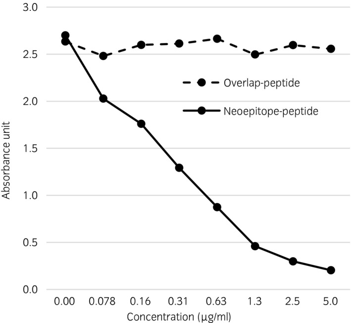
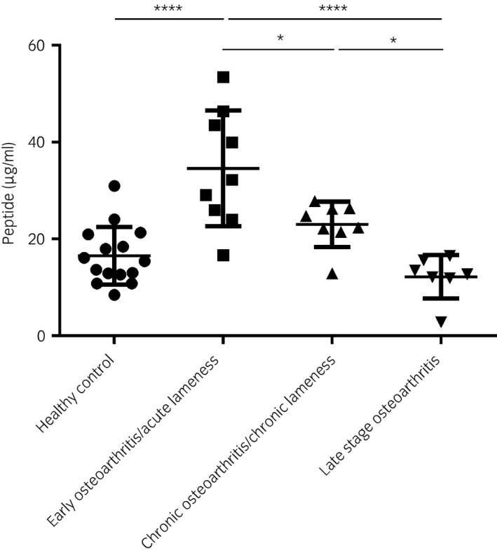
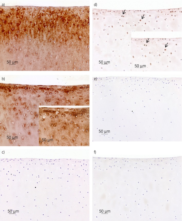
Methods: Articular cartilage explants were incubated with or without interleukin-1β for 25 days. Media were analysed via quantitative proteomics. Synovial fluid was obtained from either normal joints (n = 15) or joints causing lameness (n = 17) or with structural OA lesions (n = 7) and analysed for concentrations of the COMP neoepitope using a custom-developed inhibition enzyme-linked immunosorbent assay (ELISA). Explants were immunostained with polyclonal antibodies against COMP and the COMP neoepitopes.
Results: Semitryptic COMP peptides were identified and quantified in cell culture media from cartilage explants. A rabbit polyclonal antibody was raised against the neoepitope of the N-terminal portion of one COMP fragment (sequence SGPTHEGVC). An inhibition ELISA was developed to quantify the COMP neoepitope in synovial fluid. The mean concentration of the COMP neoepitope significantly increased in the synovial fluid from the joints responsible for acute lameness compared with normal joints and the joints of chronically lame horses and in joints with chronic structural OA. Immunolabelling for the COMP neoepitope revealed a pericellular staining in the interleukin-1β-stimulated explants.
Main limitations: The ELISA is based on polyclonal antisera rather than a monoclonal antibody.
Conclusions: The increase in the COMP neoepitope in the synovial fluid from horses with acute lameness suggests that this neoepitope has the potential to be a unique candidate biomarker for the early molecular changes in articular cartilage associated with OA.
Keywords: biomarker; cartilage oligomeric matrix protein neoepitope; horse; lameness; osteoarthritis; synovial fluid.
© 2017 The Authors Equine Veterinary Journal published by John Wiley & Sons Ltd on behalf of EVJ Ltd.
Figures
References
-
- Perkins, N.R. , Reid, S.W. and Morris, R.S. (2002) Profiling the New Zealand thoroughbred racing industry. Conditions interfering with training and racing. N.Z. Vet. J. 53, 69–76. - PubMed
-
- Secombe, C. , Firth, E. , Perkins, N. and Anderson, B. (2002) Pathophysiology and diagnosis of third carpal bone disease in horses: a review. N.Z. Vet. J. 50, 2–8. - PubMed
-
- van Weeren, P.R. and de Grauw, J. (2010) Pain in osteoarthritis. Vet. Clin. North Am. Equine Pract. 26, 619–642. - PubMed
-
- Heinegård, D. and Saxne, T. (2010) The role of the cartilage matrix in osteoarthritis. Nat. Rev. Rheumatol. 7, 50–56. - PubMed
-
- Wieland, H.A. , Michaelis, M. , Kirschbaum, B.J. and Rudolphi, K.A. (2005) Osteoarthritis – an untreatable disease? Nat. Rev. Drug Discov. 4, 331–344. - PubMed
MeSH terms
Substances
Associated data
- Actions
LinkOut - more resources
Full Text Sources
Other Literature Sources
Medical
Miscellaneous
