Cancer-associated fibroblasts promote M2 polarization of macrophages in pancreatic ductal adenocarcinoma
- PMID: 28097809
- PMCID: PMC5313646
- DOI: 10.1002/cam4.993
Cancer-associated fibroblasts promote M2 polarization of macrophages in pancreatic ductal adenocarcinoma
Abstract
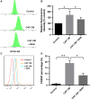
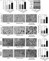
Pancreatic ductal adenocarcinoma (PDAC) is characterized by remarkable desmoplasia with infiltration of distinct cellular components. Cancer-associated fibroblasts (CAFs) has been shown to be among the most prominent cells and played a significant role in shaping the tumor microenvironment by interacting with other type of cells. Here, we aimed to investigate the effect of CAFs in modulating phenotype of tumor-associated macrophages (TAM). Under treatment of CAFs conditioned medium (CM) or direct co-culture with CAFs, monocytes exhibited enhanced expression of CD206 and CD163 compared with control group (P < 0.01). The induction of M2 polarization was mediated by increased reactive oxygen species (ROS) production in monocytes as ROS elimination abolished the effect of CAFs (P < 0.05). The supernatant analysis showed that pancreatic CAFs produced increased macrophage colony-stimulating factor (M-CSF). Upon treatment of M-CSF neutralizing antibody, the ROS generation and M2 polarization of CAFs CM-stimulated monocytes were significantly inhibited (P < 0.05). In addition, the CAFs-induced M2 macrophages significantly enhanced pancreatic tumor cell growth, migration, and invasion. Collectively, our data revealed that pancreatic CAFs were able to induce a tumor-promoting TAM phenotype partly through secreted M-CSF and enhanced ROS production in monocytes, indicating possible treatment strategies by targeting stromal cell interaction within PDAC microenvironment.
Keywords: Cancer-associated fibroblasts; Macrophage colony-stimulating factor (M-CSF); pancreatic adenocarcinoma; reactive oxygen species; tumor-associated macrophages.
© 2016 The Authors. Cancer Medicine published by John Wiley & Sons Ltd.
Figures


References
-
- Siegel, R. L. , Miller K. D., and Jemal A.. 2015. Cancer statistics, 2015. CA Cancer J. Clin. 65:5–29. - PubMed
-
- Ryan, D. P. , Hong T. S., and Bardeesy N.. 2014. Pancreatic adenocarcinoma. New England J. Med. 371:2140–2141. - PubMed
-
- Vonlaufen, A. , Phillips P. A., Xu Z., Goldstein D., R. C. Pirola , Wilson J. S., et al. 2008. Pancreatic stellate cells and pancreatic cancer cells: an unholy alliance. Cancer Res. 68:7707–7710. - PubMed
MeSH terms
Substances
LinkOut - more resources
Full Text Sources
Other Literature Sources
Medical
Research Materials

