. 2017 Feb 15;139(6):2512-2519.
doi: 10.1021/jacs.6b13399. Epub 2017 Feb 7.

Mitochondria Targeted Protein-Ruthenium Photosensitizer for Efficient Photodynamic Applications

Affiliations

Mitochondria Targeted Protein-Ruthenium Photosensitizer for Efficient Photodynamic Applications

Sabyasachi Chakrabortty et al. J Am Chem Soc. .

Abstract

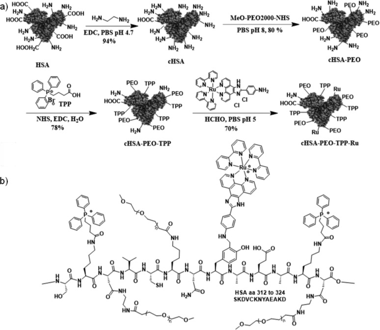
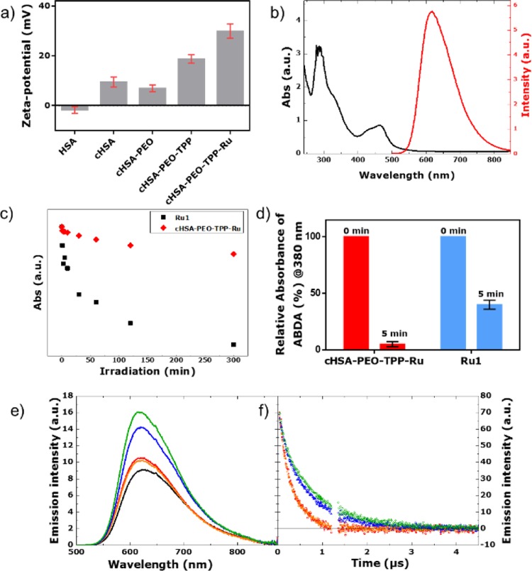
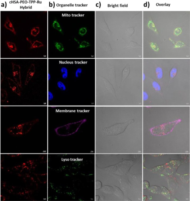
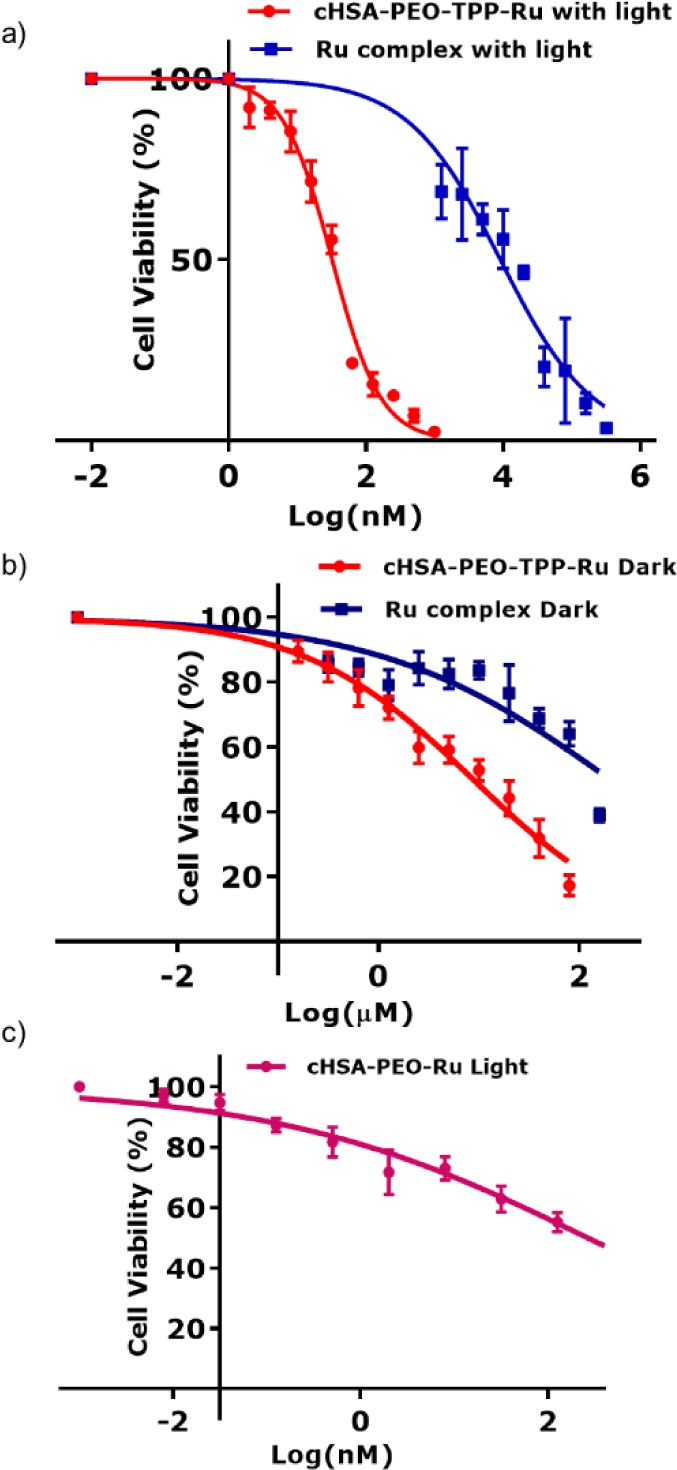
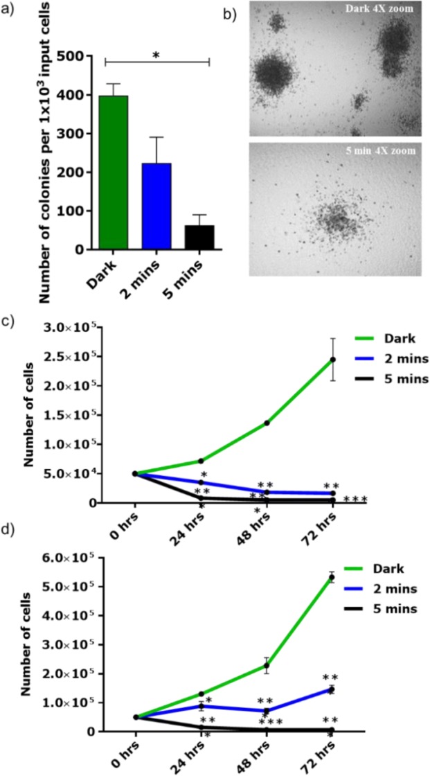
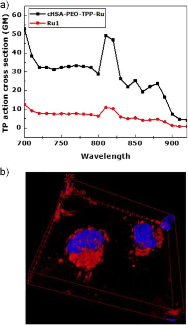
Organelle-targeted photosensitization represents a promising approach in photodynamic therapy where the design of the active photosensitizer (PS) is very crucial. In this work, we developed a macromolecular PS with multiple copies of mitochondria-targeting groups and ruthenium complexes that displays highest phototoxicity toward several cancerous cell lines. In particular, enhanced anticancer activity was demonstrated in acute myeloid leukemia cell lines, where significant impairment of proliferation and clonogenicity occurs. Finally, attractive two-photon absorbing properties further underlined the great significance of this PS for mitochondria targeted PDT applications in deep tissue cancer therapy.


Conflict of interest statement

The authors declare no competing financial interest.

Figures

Scheme 1
Scheme 1
(a) Synthetic scheme of the cHSA-PEO-TPP-Ru transporter based on HSA. Subsequent functionalities were conjugated at different reactive sites of the HSA backbone. (b) Schematic illustration of a part of the HSA polypeptide sequence exemplary with the PEO, TPP groups attached to, e.g., lysine and Ru conjugated to tyrosine residues.
Figure 1
Figure 1
(a) Zeta potential of different bioconjugates. (b) Typical absorbance and emission spectra of cHSA-PEO-TPP-Ru, where characteristic peaks of the Ru complexes are preserved in cHSA-PEO-TPP-Ru. (c) Comparison of the photostability of the Ru1 and cHSA-PEO-TPP-Ru based on the absorbance decay under continuous irradiation over extended time periods. (d) 1O2 production yield of cHSA-PEO-TPP-Ru and bare Ru complex (Ru1), as obtained from the photobleaching of the characteristic absorption peak @380 nm of ABDA (100 μM) during irradiation with 470 nm LED light (∼20 mW/cm2, 5 min) in PBS (1×, pH 7.4) based on the same optical density in their first absorption peak. (e) Steady state emission spectra (λex = 460 nm) of Ru1 complex in water (red), Ru1 complex in simulated body fluid (orange), cHSA-PEO-TPP-Ru in water (blue), cHSA-PEO-TPP-Ru in simulated body fluid (green), and Ru(bpy)3 (black) as reference at same optical density. (f) Emission lifetime experiments upon excitation at 460 nm. For the compounds, the same color code is used and an artifact region between 1.2 and 1.35 μs has been removed.
Figure 2
Figure 2
Confocal microscopy images of HeLa cells incubated with cHSA-PEO-TPP-Ru and treated with commercial organelle trackers. Overlay images and colocalization analysis of cells stained with mitochondria (0.88), nucleus (0.07), membrane (0.2), and lysosome (0.3) markers indicated that cHSA-PEO-TPP-Ru localized in mitochondria. (a) cHSA-PEO-TPP-Ru emission, (b) emission from the organelle trackers, (c) corresponding bright field images, and (d) overlay of all three images.
Figure 3
Figure 3
(a,b) Logarithmic fitting curve for cell viability of cHSA-PEO-TPP-Ru and bare Ru complex, over a broad concentration range with and without light. (c) Logarithmic fitting curve for cell viability of cHSA-PEO-Ru complex with light, where mitochondria targeting TPP group were absent. For all of the above experiments, HeLa cells were exposed to a 470 nm LED lamp (∼20 mW/cm2) for 5 min for light irradiation. cHSA-PEO-TPP-Ru reveals low dark toxicity (IC50 = 9 ± 2 μM) but very high phototoxicity (IC50 = 34.9 ± 2 nM) compared to Ru1 (dark IC50 = 203 ± 3 μM; photoirradiated IC50 = 7.7 ± 1.3 μM). In the absence of a TPP group, the phototoxic effect of the drug was reduced by ∼8 times (IC50 = 265 ± 1.2 nM).
Figure 4
Figure 4
(a) Colony forming cell (CFC) assay of the OCI-AML3 (OA3) AML cell line (n = 2 in duplicates) showed reduction of colonies after 2 and 5 min exposure to light (470 nm) compared to the treated and nonexposed cells (dark). Bars indicate mean ± SEM. Significance calculated by Mann–Whitney test (*<0.05). (b) Morphology of the colonies in the control arm and 5 min exposure arm (4× magnification). (c,d) Proliferation assay of the OA3 cell line (n = 3) incubated with two different concentrations of cHSA-PEO-TPP-Ru (75 nM) and (37.5 nM) and exposed to light for 2 and 5 min or treated but not exposed to light. Significance calculated by two-way ANOVA multiple test comparisons test (*<0.05; ***<0.0001).
Figure 5
Figure 5
(a) Two-photon absorption cross section of cHSA-PEO-TPP-Ru and Ru1 measured in water at excitation wavelengths from 700 to 920 nm using Rhodamin B as reference. (b) Two-photon 3D surface projection z-stack confocal microscopy image of HeLa cells incubated with cHSA-PEO-TPP-Ru (red, excited at 810 nm) and nuclear staining dye Hoechst (blue, excited at 405 nm).

References

    1. Ogilby P. R. Chem. Soc. Rev. 2010, 39, 3181–3209. 10.1039/b926014p. - DOI - PubMed
    1. DeRosa M. C.; Crutchley R. J. Coord. Chem. Rev. 2002, 234, 351–371. 10.1016/S0010-8545(02)00034-6. - DOI
    1. Mari C.; Pierroz V.; Ferrari S.; Gasser G. Chem. Sci. 2015, 6, 2660–2686. 10.1039/C4SC03759F. - DOI - PMC - PubMed
    1. Hamblin M. R.; Jori G.. Photodynamic Inactivation of Microbial Pathogens; Hamblin M. R., Jori G., Eds.; Comprehensive Series in Photochemical & Photobiological Sciences; The Royal Society of Chemistry: London, 2011.
    1. Wilkinson F.; Helman W. P.; Ross A. B. J. Phys. Chem. Ref. Data 1993, 22, 113–262. 10.1063/1.555934. - DOI

Publication types