. 2017 Jan 18:7:40766.
doi: 10.1038/srep40766.

Superresolution Imaging of Clinical Formalin Fixed Paraffin Embedded Breast Cancer with Single Molecule Localization Microscopy

Affiliations

Superresolution Imaging of Clinical Formalin Fixed Paraffin Embedded Breast Cancer with Single Molecule Localization Microscopy

Matthew K Creech et al. Sci Rep. .

Abstract

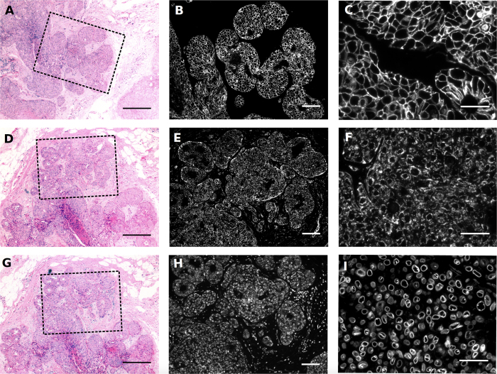
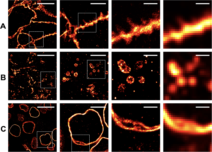
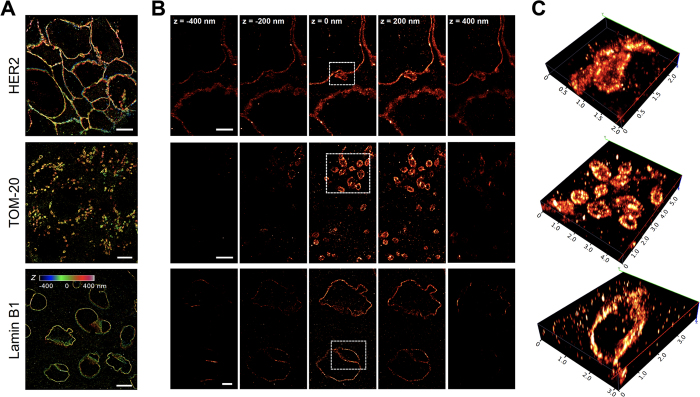
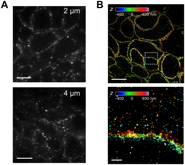
Millions of archived formalin-fixed, paraffin-embedded (FFPE) specimens contain valuable molecular insight into healthy and diseased states persevered in their native ultrastructure. To diagnose and treat diseases in tissue on the nanoscopic scale, pathology traditionally employs electron microscopy (EM), but this platform has significant limitations including cost and painstaking sample preparation. The invention of single molecule localization microscopy (SMLM) optically overcame the diffraction limit of light to resolve fluorescently labeled molecules on the nanoscale, leading to many exciting biological discoveries. However, applications of SMLM in preserved tissues has been limited. Through adaptation of the immunofluorescence workflow on FFPE sections milled at histological thickness, cellular architecture can now be visualized on the nanoscale using SMLM including individual mitochondria, undulations in the nuclear lamina, and the HER2 receptor on membrane protrusions in human breast cancer specimens. Using astigmatism imaging, these structures can also be resolved in three dimensions to a depth of ~800 nm. These results demonstrate the utility of SMLM in efficiently uncovering ultrastructural information of archived clinical samples, which may offer molecular insights into the physiopathology of tissues to assist in disease diagnosis and treatment using conventional sample preparation methods.


Figures

Figure 1
Figure 1. Morphology of FFPE HER2 positive, grade 3 IDC breast cancer tissue.
Tissue sections were stained with H&E and overview images were collected at 5X magnification to show tumor morphology (A, D and G). The box in the H&E image represents the field-of-view of the adjacent immunofluorescence images. Scale bars for H&E images = 200 μm. Images of immunofluorescence staining were collected at 10X magnification to demonstrate the tissue patterns of HER2, TOM20, and Lamin B1 proteins (B, E and H). Scale bars = 50 μm. Additional images were collected at 40X magnification to enhance visualization of ultrastructural features of each immunostain (C, F and I), scale bars = 20 μm.
Figure 2
Figure 2. Two dimensional, super resolution imaging of HER2, TOM20, and Lamin B1 in FFPE.
SMLM images at varied magnifications demonstrate the ability to resolve tissue ultrastructure not visible using conventional microscopy. Scale bars for each column of images are from left-to-right 10 μm, 4 μm, 1 μm and 1 μm, where the images in the right most column demonstrate a comparative confocal reconstruction further highlighting the necessity of SMLM to visualize nanoscopic features within the tissues. (A) Extracellular membrane protrusions were visible between HER2 positive cancer cells. (B) The outer membrane of the mitochondria and the tubular nature of this organelle were prominently resolved by SMLM imaging. (C) The lamin network of the nuclear lumen was highlighted.
Figure 3
Figure 3. Imaging HER2+ tumor FFPE sections with 3D SMLM.
(A) Reconstructed 3D SMLM images of FFPE sections immunostained for HER2 (top), TOM-20 (middle), and Lamin-B1 (bottom) with Z-positions of the molecules color coded. (B) Slice views of 3D SMLM images of HER2 (top), TOM-20 (middle), and Lamin-B1 (bottom) in 200 nm increments are demonstrated where each slice represents the projections of all localization events within 20 nm relative to the z-plane. All images were collected on 2 μm sections. (C) Volumetric rendering of the boxed areas in B are shown. Scale bars as follows: 10 μm (A), 2 μm (B, top and bottom), 1 μm (B, middle).
Figure 4
Figure 4. Comparing 2 and 4 μm FFPE sections for 3D SMLM imaging.
(A) Representative astigmatic single-molecule images collected on 2 and 4 μm sections. (B) Reconstructed 3D SMLM image of a 4 μm FFPE tumor section immunolabeled for HER2, with z-positions color coded (see legend). The bottom image shows the zoomed-in view of the boxed area in the top image. Scale bars, 5 μm (left), 10 μm (right, top) and 2 μm (right, bottom).

References

    1. Baker M. Biorepositories: Building better biobanks. Nature 486, 141–146 (2012). - PubMed
    1. Cejas P. et al. Chromatin immunoprecipitation from fixed clinical tissues reveals tumor-specific enhancer profiles. Nat Med (2016). - PubMed
    1. Lucena-Aguilar G. et al. DNA Source Selection for Downstream Applications Based on DNA Quality Indicators Analysis. Biopreserv Biobank 1–7 (2016). - PMC - PubMed
    1. Oh E. et al. Comparison of Accuracy of Whole-Exome Sequencing with Formalin-Fixed Paraffin-Embedded and Fresh Frozen Tissue Samples. PLoS One 10, e0144162 (2015). - PMC - PubMed
    1. Najarian R. et al. Primary structure and gene organization of human hepatitis A virus. Proc Natl Acad Sci USA 82, 2627–2631 (1985). - PMC - PubMed

Publication types

Substances