. 2017 Jan 18:7:40753.
doi: 10.1038/srep40753.

Vanillic acid attenuates Aβ1-42-induced oxidative stress and cognitive impairment in mice

Affiliations

Vanillic acid attenuates Aβ1-42-induced oxidative stress and cognitive impairment in mice

Faiz Ul Amin et al. Sci Rep. .

Abstract

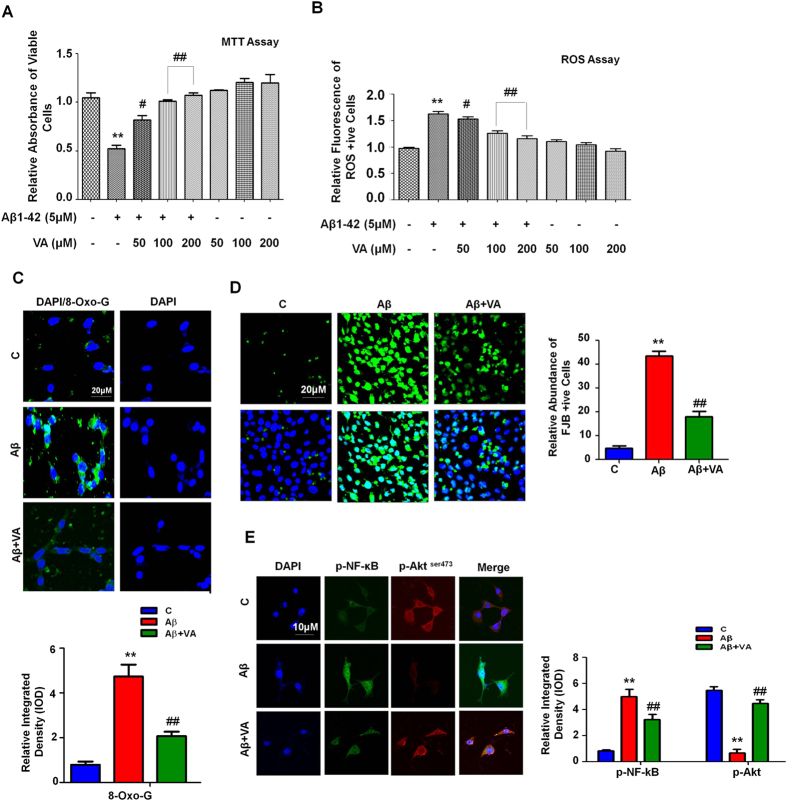
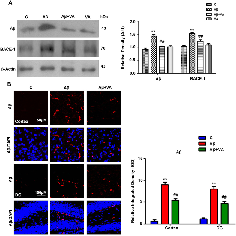
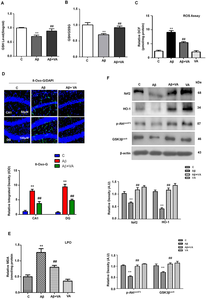
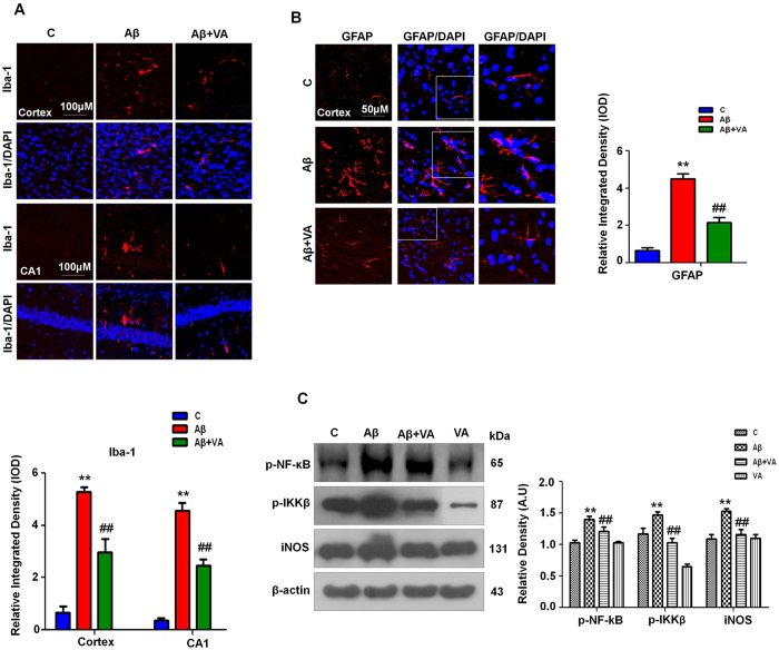
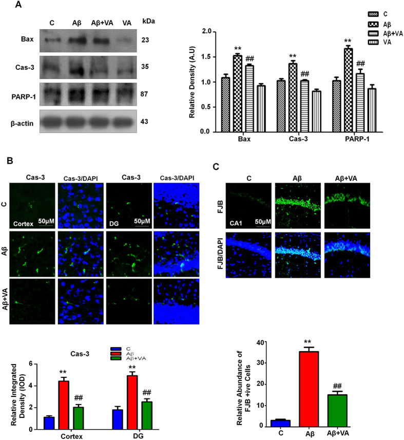
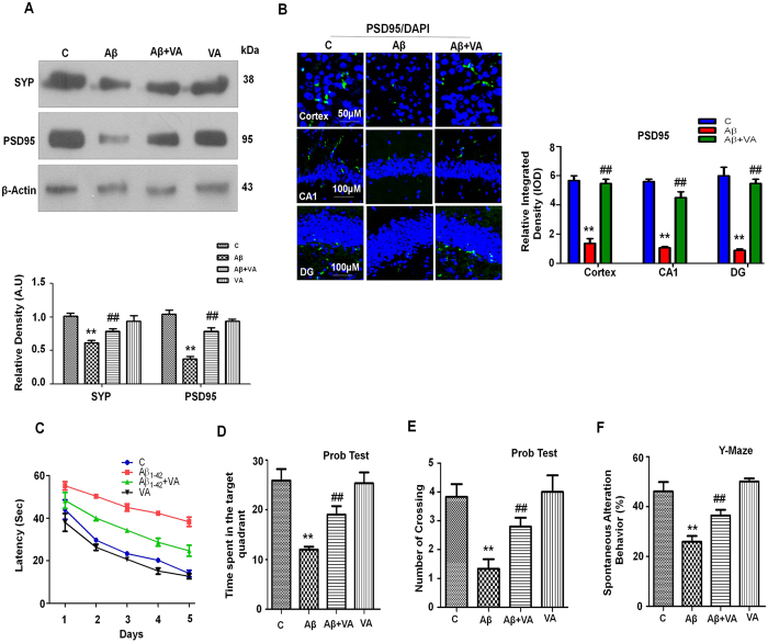
Increasing evidence demonstrates that β-amyloid (Aβ) elicits oxidative stress, which contributes to the pathogenesis and disease progression of Alzheimer's disease (AD). The aims of the present study were to determine and explore the antioxidant nature and potential mechanism of vanillic acid (VA) in Aβ1-42-induced oxidative stress and neuroinflammation mediated cognitive impairment in mice. An intracerebroventricular (i.c.v.) injection of Aβ1-42 into the mouse brain triggered increased reactive oxygen species (ROS) levels, neuroinflammation, synaptic deficits, memory impairment, and neurodegeneration. In contrast, the i.p. (intraperitoneal) administration of VA (30 mg/kg, for 3 weeks) after Aβ1-42-injection enhanced glutathione levels (GSH) and abrogated ROS generation accompanied by an induction of the endogenous nuclear factor erythroid 2-related factor 2 (Nrf2) and heme oxygenase 1 (HO-1) via the activation of Akt and glycogen synthase kinase 3β (GSK-3β) in the brain mice. Additionally, VA treatment decreased Aβ1-42-induced neuronal apoptosis and neuroinflammation and improved synaptic and cognitive deficits. Moreover, VA was nontoxic to HT22 cells and increased cell viability after Aβ1-42 exposure. To our knowledge, this study is the first to reveal the neuroprotective effect of VA against Aβ1-42-induced neurotoxicity. Our findings demonstrate that VA could potentially serve as a novel, promising, and accessible neuroprotective agent against progressive neurodegenerative diseases such as AD.


Figures

Figure 1
Figure 1. The beneficial effects of VA on Aβ1-42-induced neurotoxicity in vitro.
(A) Shown is the cell viability (MTT assay) histogram. Aβ1-42 (5 μM) reduced cell viability; treatment with VA at three different concentrations (50, 100 and 200 μM) increased the viability of HT22 cells after 24 h. (B) Representative ROS assay histogram. Treatment with vanillic acid at all three different concentrations (50, 100 and 200 μM) significantly reduced Aβ1-42-induced (5 μM) ROS levels. These assays were performed in triplicate (±S.E.M.). (C) The immunofluorescence images along with their respective histogram of 8-OxoG (green) counterstained with DAPI (blue) in all three treated groups in HT22 cells. (D) Shown are the immunofluorescence images of FJB staining along with the respective integrated density histograms in HT22 cells treated with Aβ1-42 (5 μM) and VA (100 μM) for 24 h. (E) The double immunofluorescence images of HT22 cells after Aβ1-42 and VA treatment for 24 h, showing p-NF-kB (green), p-Akt (red), proteins and their respective relative density histograms. DAPI (blue) was used to counterstain the nucleus. These experiments were performed in triplicate. Details are given in the methods section. *Significantly different from vehicle-treated animals; #significantly different from Aβ1-42-treated animals. Significance = **P < 0.01, #P < 0.05, ##P < 0.01.
Figure 2
Figure 2. Vanillic acid attenuated Aβ accumulation and β-site APP cleaving enzyme 1 (BACE-1) overexpression in mouse brain homogenates.
(A) Immunoblot analysis of Aβ and BACE- 1 protein expression in the mouse brain following Aβ and VA administration. The bands were quantified using Sigma Gel software, and the differences are represented by a histogram. β-Actin was used as a loading control. The density values are expressed in arbitrary units (A.U.) as the mean ± S.E.M. for the indicated protein (n ± 5 mice/group). (B) The immunofluorescence of Aβ was used to evaluate the cortex and hippocampus of experimental mice (n ± 8 mice/group). Magnification, 10X. *Significantly different from vehicle-treated animals; #significantly different from Aβ1-42-treated animals. Significance  ± **P < 0.01; ##P < 0.01.
Figure 3
Figure 3. Vanillic acid treatment ameliorates ROS and oxidative stress in Aβ1-42-treated mice.
(A) A representative histogram showing the ROS level in the mouse brain (n ± 5 mice/group). (B) A representative histogram showing GSH levels in the brains of mice. GSH levels were measured with a colorimetric assay kit and were expressed as nmol/mg protein. (C) A representative histogram showing the GSH/GSSG levels in the brains of mice. (D) The images given shows the immunoreactivity of 8-OxoG (green) along with their respective histogram counterstained with DAPI (blue) in all three treated groups in the hippocampal CA1 and DG region. (E) The histogram depicts the LPO levels in the treated groups. The methods details are given in the material and methods section. All these experiments were performed in triplicates. (F) Vanillic acid treatment stimulated the Akt/GSK3β/Nrf2/HO-1 pathway in the brains of Aβ1-42-treated mice. Western blot analysis demonstrated the expression of Nrf2, HO-1, p-Akt, and GSK3β in the brains of mice. The bands were quantified using Sigma Gel software, and the differences are represented in a histogram. β-Actin was used as a loading control. The density values are expressed in arbitrary units (A.U.) as the mean ± S.E.M. for the indicated proteins (n ± 5 mice/group). *Significantly different from vehicle-treated mice; #significantly different from Aβ1-42-treated mice. Significance = **P < 0.01; ##P < 0.01.
Figure 4
Figure 4. Vanillic acid treatment attenuated the number of Aβ1-42-induced activated glial cells (microglia and astrocytes) and reduced neuroinflammation in the mouse brain (n ± 8 mice/group).
(A) Iba-1 immunofluorescence revealed a significant increase in the number of Iba-1 reactive cells in the brains of mice in the Aβ1-42-treated group compared to mice in the vehicle-treated group. On the other hand, VA treatment significantly decreased the number of reactive Iba-1 cells in the brains of mice exposed to Aβ1-42. (B) Immunofluorescence images revealed a significant increase in the number of GFAP reactive cells in the brains of mice in the Aβ1-42-treated group compared to the vehicle-treated group. Vanillic acid treatment significantly decreased the number of reactive GFAP cells in the brains of mice exposed to Aβ1-42. (C) Western blot analysis of p-IKKβ, p-NF-κB and iNOS in the brain of mice. The bands were quantified using Sigma Gel software, and the differences are represented in a histogram. β-Actin was used as a loading control. The density values are expressed in arbitrary units (A.U.) as the mean ± SEM for the indicated proteins (n ± 5 mice/group). *Significantly different from vehicle-treated mice; #significantly different from Aβ1-42-treated mice. Significance = **P < 0.01, ##P < 0.01.
Figure 5
Figure 5. Vanillic acid prevented Aβ1-42-induced apoptosis and neurodegeneration.
(A) Immunoblot analysis of the mouse brain using activated Bax, caspase-3 and cleaved PARP-1 antibodies. The bands were quantified using Sigma Gel software, and the differences are represented in a histogram. Anti-β-actin was used as a loading control. The density values are expressed in arbitrary units (A.U.) as the mean ± S.E.M. for the indicated brain proteins (n ± 5 mice/group). (B) Immunofluorescence of activated caspase-3 in the cortex and hippocampus in the experimental mice (n ± 8 mice/group). Caspase-3-positive neurons were increased in the Aβ1-42-treated mice compared with the control mice. Vanillic acid treatment significantly decreased the number of Aβ1-42-induced caspase-3-positive neurons. (C) Immunofluorescence of FJB positive neurons in the CA1 region of vehicle, Aβ1-42 and VA treated mouse brains. Magnification, 10X. *Significantly different from the vehicle-treated; #significantly different from Aβ1-42 treated. Significance = **P < 0.01 and ##P < 0.01.
Figure 6
Figure 6. Vanillic acid treatment attenuated Aβ1-42-induced synaptic disorganization and cognitive impairment in mice.
(A) Immunoblot analysis of synaptophysin and PSD95 in the mouse brain. The bands were quantified using Sigma Gel software, and the differences are represented in a histogram. β-Actin was used as a loading control. The density values are expressed in arbitrary units (A.U.) as the mean ± S.E.M. for the indicated proteins (n ± 5 mice/group). (B) A representative immunofluorescence image of PSD95 reactivity in the cortex and hippocampus of experimental mice (n ± 8 mice/group). Magnification, 10X. (C) A representative histogram for the mean escape latency (sec) in the MWM test during the training session (n ± 13 mice/group). (D) Time spent in the target quadrant during the probe test. (E) The number of platform crossings during the probe test. (F) A histogram showing the percentage spontaneous alterations in the Y-maze test (n = 13 mice/group). *Significantly different from the vehicle-treated mice; #significantly different from Aβ1-42 treated mice. Significance = **P < 0.01, ##P < 0.01.

References

    1. Lambracht-Washington D. & Rosenberg R. N. Advances in the development of vaccines for Alzheimer’s disease. Discov Med. 15, 319–326 (2013). - PMC - PubMed
    1. Coomaraswamy J. et al. Modeling familial Danish dementia in mice supports the concept of the amyloid hypothesis of Alzheimer’s disease. Proc Natl Acad Sci USA 107, 7969–7974 (2010). - PMC - PubMed
    1. Chan K. H. et al. Adiponectin is protective against Oxidative stress induced cytotoxicity in Amyloid-beta neurotoxicity. PLoS ONE 7, 52354 (2012). - PMC - PubMed
    1. Lamert M. P. et al. Diffusable, nonfibrillar ligands derived from Ab1-42 are potent central nervous system neurotoxins. Proc Natl Acad Sci USA 95, 6448–6453 (1998). - PMC - PubMed
    1. Jhoo J. H. et al. Beta-Amyloid (1-42)-induced learning and memory deficits in mice: involvement of oxidative burdens in the hippocampus and cerebral cortex. Behav Brain Res 155, 185–196 (2004). - PubMed

Publication types

MeSH terms