. 2017 Jan 18:7:40783.
doi: 10.1038/srep40783.

Hepatitis B virus X protein is capable of down-regulating protein level of host antiviral protein APOBEC3G

Affiliations

Hepatitis B virus X protein is capable of down-regulating protein level of host antiviral protein APOBEC3G

Ruidong Chen et al. Sci Rep. .

Abstract

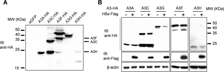
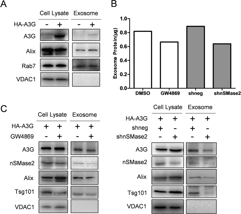
The apolipoprotein B mRNA editing catalytic polypeptide-like (APOBEC) family proteins bind RNA and single-stranded DNA, and create C-to-U base modifications through cytidine deaminase activity. APOBEC3G restricts human immunodeficiency virus 1 (HIV-1) infection by creating hypermutations in proviral DNA, while HIV-1-encoded vif protein antagonizes such restriction by targeting APOBEC3G for degradation. APOBEC3G also inhibits hepatitis B virus (HBV): APOBEC3G co-expression inhibits HBV replication and evidences exist indicating APOBEC3G-mediated HBV hypermutations in patients. HBV encodes a small non-structural X protein (HBx) with a recognized activating effect on HBV life cycle. In this work, we report the discovery that HBx selectively and dose-dependently decreases the protein level of co-expressed APOBEC3G in transfected Huh-7 cells. The effect was shown to take place post-translationally, but does not rely on protein degradation via proteasome or lysosome. Further work demonstrated that intracellular APOBEC3G is normally exported via exosome secretion and inhibition of exosome biogenesis causes retention of intracellular APOBEC3G. Finally, HBx co-expression specifically enhanced externalization of APOBEC3G via exosomes, resulting in decrease of intracellular APOBEC3G protein level. These data suggest the possibility that in addition to other mechanisms, HBx-mediated activation of HBV might also involve antagonizing of intracellular restriction factor APOBEC3G through promotion of its export.


Figures

Figure 1
Figure 1. HBx selectively decreases A3G protein level.
(A) Confirmation of expression of selected APOBEC3 (A3) family members. Huh-7 cells were transfected with expression plasmids encoding HA-tagged A3 proteins or eGFP control plasmid as indicated. Cells were harvested 48 hours post transfection and cell lysates were analyzed in parallel using immunoblot. Arrows indicate positions of full-length A3C, A3F and A3H. (B) Effects of HBx co-transfection on A3 protein levels. Huh-7 cells were transfected with indicated A3 member plasmids with or without HBx expression plasmid. Cells were harvested 48 hours post transfection. A3 proteins, HBx and β-actin were detected using antibodies as indicated.
Figure 2
Figure 2. HBx dose-dependently decreases A3G protein level.
(A) Huh-7 cells were transfected with A3G and increasing amounts of HBx as indicated. eGFP expression plasmid was used as transfection control. Cells were harvested 48 hours post transfection and cell lysates were analyzed in parallel in immunoblot. (B) Relative quantities of A3G in cell lysates as shown in (A) were estimated using densitometry scanning and normalized to eGFP quantities, taking values without HBx co-transfection as 1 (100%). Means and SD calculated from 3 independent experiments are shown.
Figure 3
Figure 3. HBx decreases A3G protein level post-translationally.
(A) Time-course study of A3G protein level following cycloheximide (CHX) treatment. Huh-7 cells transfected with A3G and eGFP expression plasmids, with or without HBx plasmid, were treated with CHX at 48 hours post transfection. Cells were collected at indicated time points and analyzed in parallel using immunoblot. (B) Relative quantities of A3G (top) and HBx (bottom) in cell lysates as shown in (A) were estimated using densitometry scanning and normalized against eGFP quantities, taking values at start of CHX treatment as 1 (100%). Means and SD calculated from 3 independent experiments are shown.
Figure 4
Figure 4. Degradation of A3G is not markedly affected by HBx.
(A) Lack of effects of HBx on A3G ubiquitination as detected using immunoprecipitation. Huh-7 cells transfected with indicated plasmids were treated with MG-132 and subjected to cell lysis and purification of His-tagged A3G using Ni2+-NTA matrix under denaturing condition. Enriched His-tagged A3G was detected using anti-A3G and A3G modified by HA-tagged ubiquitin (HA-Ub) was detected using anti-HA antibodies. A3G and HBx in 1/10 input were also detected. (B and C) Treatment with proteasome inhibitor MG-132 or lysosome inhibitor leupeptin had no marked effect on A3G protein levels, with or without HBx. Huh-7 cells transfected and treated as indicated were lysed and subjected to immunoblot analysis.
Figure 5
Figure 5. Intracellular A3G is exported via exosome secretion.
(A) Detection of A3G protein in cell lysates and exosomes prepared from Huh-7 cells transfected with A3G expression plasmid or vector control. Exosome markers Alix and Rab7, and mitochondria marker VDAC1 were detected in parallel. (B) Treatment with GW4869 or transfection of plasmid expressing nSMase2-targeting shRNA (shnSMase2) inhibited exosome secretion by Huh-7 cells. Exosomes were prepared from 1E7 cells treated as indicated and total protein contents were quantified using BCA assay. DMSO vehicle and unrelated shRNA construct (shneg) were used as controls. (C) Treatment with GW4869 or shnSMase2 inhibited A3G export via exosome secretion and increased intracellular A3G protein level. Huh-7 cells transfected with A3G expression plasmid or vector control and treated as indicated were used for lysate and exosome preparation. In addition to A3G and exosome/mitochondria markers, nSMase2 was also detected to demonstrate knock-down by shnSMase2.
Figure 6
Figure 6. HBx increases export of A3G via exosome secretion.
(A) Effects of HBx on A3G protein levels in cell lysates and exosomes. Huh-7 cells were transfected with indicated plasmids and 48 hours post transfection, cell lysates and exosomes were prepared and analyzed in immunoblot for A3G and HBx levels as well as exosome markers Tsg101 and Rab7. β-actin was used as loading control. (B) Relative quantities of A3G and exosome markers Tsg101 and Rab7 in cell lysates and exosomes as shown in (A) were respectively estimated using densitometry scanning taking values from vector-transfected cells as 1 (100%). Means and SD calculated from 3 independent experiments are shown.

Similar articles

Cited by

References

    1. Seeger C. & Mason W. S. Hepatitis B virus biology. Microbiol Mol Biol Rev 64, 51–68 (2000). - PMC - PubMed
    1. Seeger C. & Mason W. S. Molecular biology of hepatitis B virus infection. Virology 479–480, 672–686, doi: 10.1016/j.virol.2015.02.031 (2015). - DOI - PMC - PubMed
    1. Liang T. J. Hepatitis B: the virus and disease. Hepatology 49, S13–21, doi: 10.1002/hep.22881 (2009). - DOI - PMC - PubMed
    1. World Health Organization. Hepatitis B. World Health Organization Fact Sheet 204 (Revised July 2016). http://www.who.int/mediacentre/factsheets/fs204/en/ (2016).
    1. Protzer U. Hepatitis: Epigenetic control of HBV by HBx protein–releasing the break? Nature reviews. Gastroenterology & hepatology 12, 558–559, doi: 10.1038/nrgastro.2015.152 (2015). - DOI - PubMed

Publication types

MeSH terms

Substances