

Review
doi: 10.1249/JES.0000000000000101.
Mitochondria Initiate and Regulate Sarcopenia
Affiliations
- PMID: 28098577
- PMCID: PMC5357179
- DOI: 10.1249/JES.0000000000000101
Item in Clipboard
Review
Mitochondria Initiate and Regulate Sarcopenia
Exerc Sport Sci Rev.
2017 Apr.
Abstract
We present the hypothesis that an accumulation of dysfunctional mitochondria initiates a signaling cascade leading to motor neuron and muscle fiber death and culminating in sarcopenia. Interactions between neural and muscle cells that contain dysfunctional mitochondria exacerbate sarcopenia. Preventing sarcopenia will require identifying mitochondrial sources of dysfunction that are reversible.
Figures
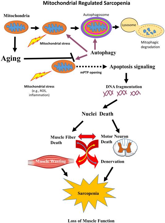
Induction of mitochondrial stress (lightning bolt) can result in dysfunctional mitochondria. Damaged mitochondria are engulfed in an autophagosome membrane and removed by mitophagy signaling in healthy young muscle and motor neurons. However, aging is associated with increased ROS and other mitochondrial stresses, which enhance mPTP opening. Release of the mitochondrial contents to the cell cytosol induces an apoptotic cascade ending with DNA fragmentation and removal of nuclei. Sufficient nuclear death in muscle cells will result in the death and removal of the entire muscle cell. Similarly, motor neuron death occurs when apoptosis removal of the alpha motor neuron nucleus occurs. The interdependence of muscle cells and motor neurons suggests a potential feedback loop (anterograde and retrograde) communication between the muscle and the motor neuronal compartments, which exacerbates death in both compartments. Death in these cell compartments leads to loss of muscle mass and function in aging. Thus, dysfunctional mitochondria provide the signal to initiate sarcopenia.
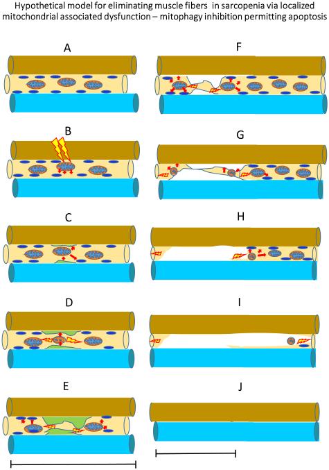
A. In healthy muscle, activation of mitophagy eliminates dysfunctional nuclei so that they cannot continue death signaling. B. Dysfunctional mitochondria that leak their contents to the cytosol will occur in muscle that has received a significant mitochondrial stress (e.g., ROS, inflammatory mediators etc.). C-D. This initiates the apoptotic signaling cascades. E-F. If the dysfunctional mitochondria are not eliminated, apoptotic death signaling may be activated to eliminate myonuclei and this may concurrently or independently result in wide-spread activation of autophagy and the ubiquitin ligase pathway and also, trigger the necrosis signaling pathway to remove muscle proteins, mitochondria and nuclei within the domain of the initial dysfunctional mitochondria (G-H). I-J. The extent of dysfunctional mitochondrial will extend along the mitochondrial reticular network and affect the function of other mitochondria near the dysfunctional mitochondrial. The wider accumulation of dysfunctional mitochondria will perpetuate signaling for apoptosis, which will remove nuclei from a larger area. The greater nuclear loss will be followed by elevated proteasome signaling to eliminate contractile and non-contractile tissue in the fiber segment that is associated with dysfunctional mitochondria and apoptotic signaling. This cellular removal will result in eventual elimination of the portion of the fiber in the area of the dysfunctional mitochondrial and potentially the entire fiber. We further hypothesize that the initiation of the disassembly and removal of the fiber could be blocked if the dysfunctional mitochondria which initiate the process, are removed or the damage to mitochondria reversed (e.g., via exercise and nutritional interventions) and if irreparable damaged mitochondria are replaced by healthy mitochondria.
References
-
- Adhihetty PJ, O'Leary MF, Chabi B, Wicks KL, Hood DA. Effect of denervation on mitochondrially mediated apoptosis in skeletal muscle. J. Appl. Physiol. 2007;102:1143–1151. - PubMed
-
- Aiken J, Bua E, Cao Z, Lopez M, Wanagat J, McKenzie D, McKiernan S. Mitochondrial DNA deletion mutations and sarcopenia. Ann. N. Y. Acad. Sci. 2002;959:412–423. - PubMed
-
- Alway SE, Degens H, Krishnamurthy G, Chaudhrai A. Denervation stimulates apoptosis but not Id2 expression in hindlimb muscles of aged rats. J Gerontol. A Biol. Sci Med Sci. 2003;58:687–697. - PubMed
Publication types
MeSH terms
Substances
Grants and funding
LinkOut - more resources
Full Text Sources
Other Literature Sources
Medical
