. 2017 Jan 13;22(1):126.
doi: 10.3390/molecules22010126.

Penduliflaworosin, a Diterpenoid from Croton crassifolius, Exerts Anti-Angiogenic Effect via VEGF Receptor-2 Signaling Pathway

Affiliations

Penduliflaworosin, a Diterpenoid from Croton crassifolius, Exerts Anti-Angiogenic Effect via VEGF Receptor-2 Signaling Pathway

Yeyin Liang et al. Molecules. .

Abstract

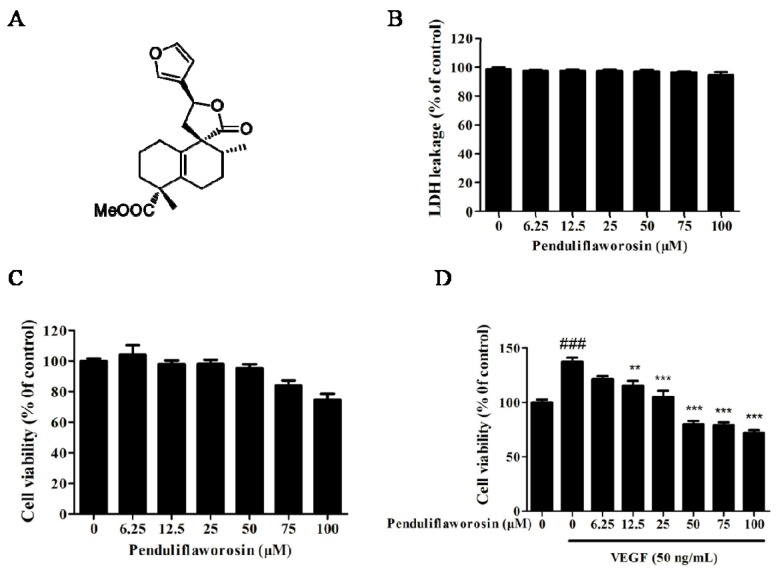
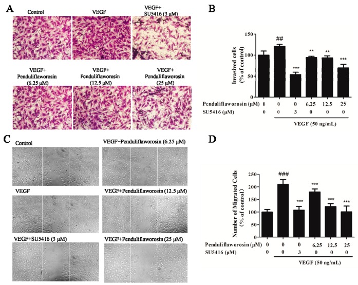
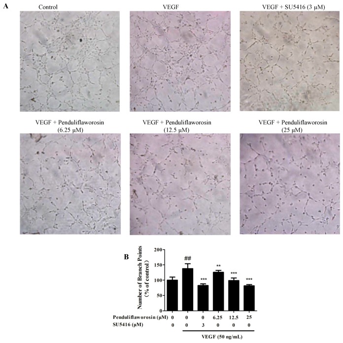
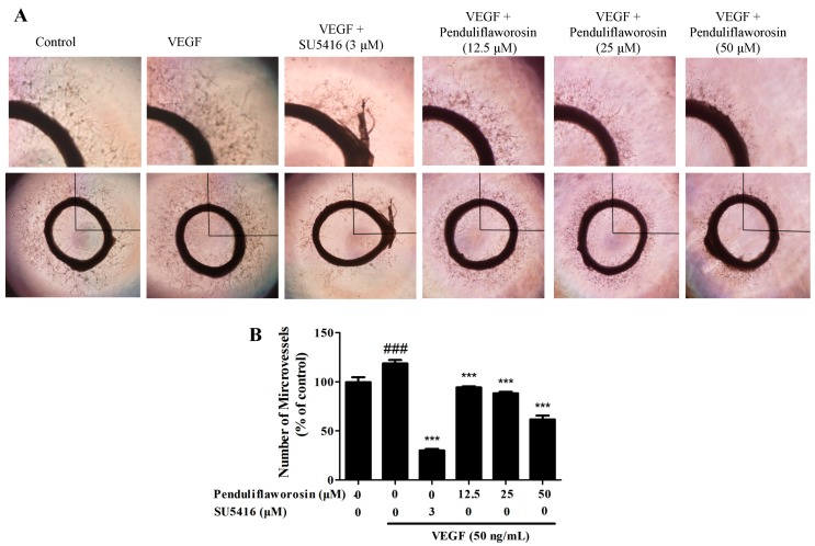
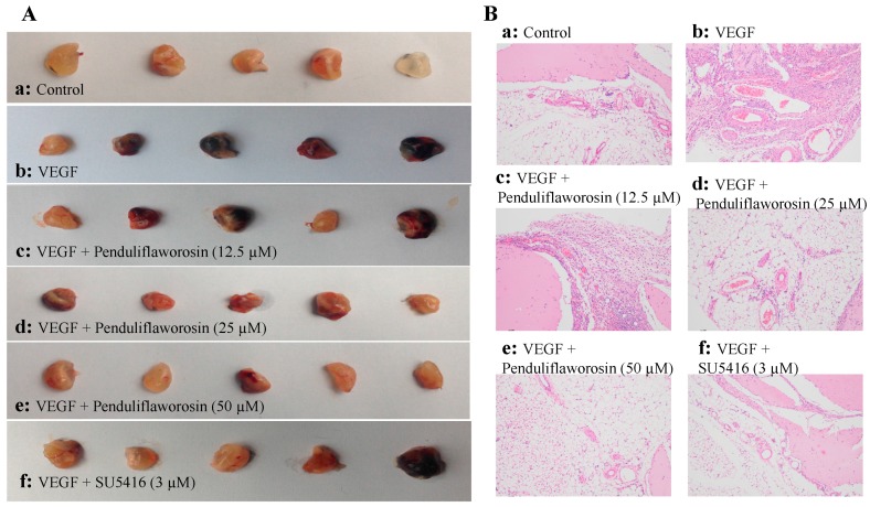
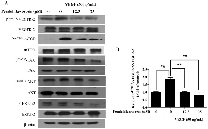
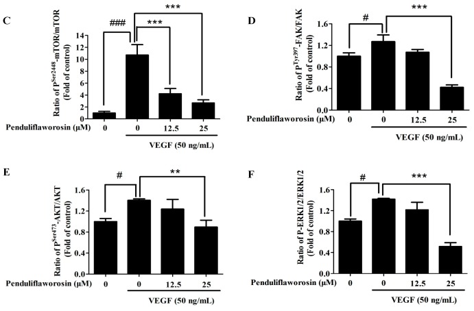
Anti-angiogenesis targeting vascular endothelial growth factor receptor-2 (VEGFR-2) has been considered as an important strategy for cancer therapy. Penduliflaworosin is a diterpenoid isolated from the plant Croton crassifolius. Our previous study showed that this diterpenoid possesses strong anti-angiogenic activity by inhibiting vessel formation in zebrafish. This study was conducted to further investigate the anti-angiogenic activity and mechanism of penduliflaworosin. Results revealed that penduliflaworosin significantly inhibited VEGF-induced angiogenesis processes including proliferation, invasion, migration, and tube formation of human umbilical vein endothelial cells (HUVECs). Moreover, it notably inhibited VEGF-induced sprout formation of aortic rings and blocked VEGF-induced vessel formation in mice. Western blotting studies showed that penduliflaworosin inhibited phosphorylation of the VEGF receptor-2 and its downstream signaling mediators in HUVECs, suggesting that the anti-angiogenic activity was due to an interference with the VEGF/VEGF receptor-2 pathway. In addition, molecular docking simulation indicated that penduliflaworosin could form hydrogen bonds within the ATP-binding region of the VEGF receptor-2 kinase unit. Finally, cytotoxicity assay showed that penduliflaworosin possessed little toxicity toward both cancer and normal cells. Taken together, our findings demonstrate that penduliflaworosin exerts its anti-angiogenic effect via the VEGF receptor-2 signaling pathway. The anti-angiogenic property and low cytotoxicity of penduliflaworosin suggest that it may be useful in cancer treatments.

Keywords: VEGF receptor-2; anti-angiogenesis; penduliflaworosin.


Conflict of interest statement

The authors declare no conflict of interest.

Figures

Figure 1
Figure 1
(A) The structure of penduliflaworosin. Cytotoxicity of penduliflaworosin in human umbilical vein endothelial cells (HUVECs) was evaluated by (B) lactate dehydrogenase (LDH) assay and (C) cell-counting assay; (D) Penduliflaworosin inhibited vascular endothelial growth factor (VEGF)-induced proliferation of HUVECs in a dose-dependent manner at 48 h. Cells treated with medium only were used as the vehicle control, and data are shown as a percentage of the control. Data are expressed as the mean ± standard deviation (n = 3). Mean values showing significant differences between the control group and the VEGF-treated group are indicated by ### (p < 0.001). Mean values showing significant difference between the VEGF-treated group and the penduliflaworosin-treated group are denoted by ** (p < 0.01) or *** (p < 0.001).
Figure 2
Figure 2
Penduliflaworosin suppressed VEGF-induced invasion and migration of HUVECs. The (A) invasion and (C) migration of HUVECs were photographed. The numbers of (B) invading and (D) migrating cells were counted after treatment with compounds (penduliflaworosin or SU5416) at different concentrations. Cells treated with medium only were used as the vehicle control. Data are shown as the percentage of the vehicle-treated control. Values are represented as the mean ± standard deviation (n = 3). Mean values showing significant differences between the control group and the VEGF-treated group are denoted by ## (p < 0.05) or ### (p < 0.001). Mean values showing significant differences between the VEGF-treated group and the penduliflaworosin-treated group are indicated by ** (p < 0.01), or *** (p < 0.001).
Figure 3
Figure 3
Penduliflaworosin suppressed VEGF-induced tube formation of HUVECs in a dose-dependent manner. (A) The tube formation of HUVECs was photographed. The numbers of (B) branch points in the tubes were counted after treatment with different concentrations of compounds (penduliflaworosin or SU5416). Cells with medium only were used as the vehicle control, and data are presented as the percentage of the control. Values are represented as the mean ± standard deviation (n = 3). Mean values showing significant differences between the control group and the VEGF-treated group are denoted by ## (p < 0.001). Mean values showing significant difference between the VEGF-treated group and penduliflaworosin-treated group are indicated by ** (p < 0.01) or *** (p < 0.001).
Figure 4
Figure 4
Penduliflaworosin inhibited VEGF-induced vessel sprout formation in a dose-dependent manner. Aortic rings isolated from Sprague-Dawley rats were cultured in wells pre-coated with Matrigel and treated with different concentrations of compounds (penduliflaworosin or SU5416) and VEGF (50 ng/mL) for 4 days. Aortic rings treated with culture medium only were used as the vehicle control. (A) The sprouting of microvessels from the aortic rings was photographed; (B) The numbers of microvessels sprouting from aortic rings were counted after treatment with the compounds. Aortic rings treated with medium only were used as the vehicle control, and data are shown as a percentage of the control. Values are represented as the mean ± standard deviation (n = 3). Mean values showing significant differences between the control group and the VEGF-treated group are denoted by ### (p < 0.001). Mean values showing significant difference between the VEGF-treated group and penduliflaworosin-treated group are indicated by *** (p < 0.001).
Figure 5
Figure 5
Penduliflaworosin inhibited VEGF-induced blood vessel formation in vivo. The ventral area of C57/BL/6 mice was injected with 300 µL of Matrigel containing various concentrations of compounds in presence of VEGF (250 ng) and heparin (150 units). After incubation for 21 days, the Matrigel plugs were harvested. (A) The Matrigel plugs were photographed; (B) the Matrigel plugs were fixed, sectioned, and stained with hematoxylin and eosin (magnification, ×200).
Figure 6
Figure 6
(A) Effect of penduliflaworosin on the expression of the phosphorylation of VEGF receptor-2 (VEGFR-2) and VEGFR-2 downstream signaling molecules, including mammalian target of rapamycin (mTOR), focal adhesion kinase (FAK), protein kinase B (AKT), and extracellular signal-regulated kinase (ERK) in HUVECs in a dose-dependent manner. (BF) The ratios of phosphoTyr1175-VEGFR-2/VEGFR-2, phosphoSer2448-mTOR/mTOR, phosphoTyr397-FAK/FAK, phosphoSer473-AKT/AKT, and phospho-ERK1/2/ERK1/2 were determined, respectively. Values are represented as the mean ± standard deviation (n = 3). Mean values showing significant differences between the control group and the VEGF-treated group are denoted by # (p < 0.05), ## (p < 0.01), or ### (p < 0.001). Mean values showing significant difference between the VEGF-treated group and penduliflaworosin-treated group are indicated by ** (p < 0.01) or *** (p < 0.001).
Figure 6
Figure 6
(A) Effect of penduliflaworosin on the expression of the phosphorylation of VEGF receptor-2 (VEGFR-2) and VEGFR-2 downstream signaling molecules, including mammalian target of rapamycin (mTOR), focal adhesion kinase (FAK), protein kinase B (AKT), and extracellular signal-regulated kinase (ERK) in HUVECs in a dose-dependent manner. (BF) The ratios of phosphoTyr1175-VEGFR-2/VEGFR-2, phosphoSer2448-mTOR/mTOR, phosphoTyr397-FAK/FAK, phosphoSer473-AKT/AKT, and phospho-ERK1/2/ERK1/2 were determined, respectively. Values are represented as the mean ± standard deviation (n = 3). Mean values showing significant differences between the control group and the VEGF-treated group are denoted by # (p < 0.05), ## (p < 0.01), or ### (p < 0.001). Mean values showing significant difference between the VEGF-treated group and penduliflaworosin-treated group are indicated by ** (p < 0.01) or *** (p < 0.001).
Figure 7
Figure 7
Penduliflaworosin interacted with the ATP-binding sites of VEGF receptor-2 kinase domain. (A) Penduliflaworosin formed hydrogen bonds with residue Asp1046. (B) MOLCAD surface modeling showed that the furan ring of penduliflaworosin extended into the deep cavity of the ATP-binding pocket of VEGF receptor-2.

Similar articles

Cited by

References

    1. Folkman J. Angiogenesis in cancer, vascular, rheumatoid and other disease. Nat. Med. 1995;1:27–31. doi: 10.1038/nm0195-27. - DOI - PubMed
    1. Michael P., Herman I.M. Mechanisms of normal and tumor-derived angiogenesis. Am. J. Physiol.-Cell. Physiol. 2002;282:C947–C970. - PubMed
    1. David B., Harold W. Targeting the growth factors and angiogenesis pathways: Small molecules in solid tumors. J. Surg. Oncol. 2011;103:574–586. - PubMed
    1. Carmeliet P., Jain R.K. Molecular mechanisms and clinical applications of angiogenesis. Nature. 2011;473:298–307. doi: 10.1038/nature10144. - DOI - PMC - PubMed
    1. Fyfe G.A., Hurwitz H., Fehrenbacher L., Cartwright T., Hainsworth J., Heim W., Berlin J., Kabbinavar F., Holmgren E., Novotny W. Bevacizumab plus irinotecan/5-FU/leucovorin for treatment of metastatic colorectal cancer results in survival benefit in all pre-specified patient subgroups. J. Clin. Oncol. 2004;22:3617.

MeSH terms

Substances

LinkOut - more resources