

Lymphangioleiomyomatosis: A Monogenic Model of Malignancy
- PMID: 28099079
- PMCID: PMC5663315
- DOI: 10.1146/annurev-med-050715-104245
Lymphangioleiomyomatosis: A Monogenic Model of Malignancy
Abstract
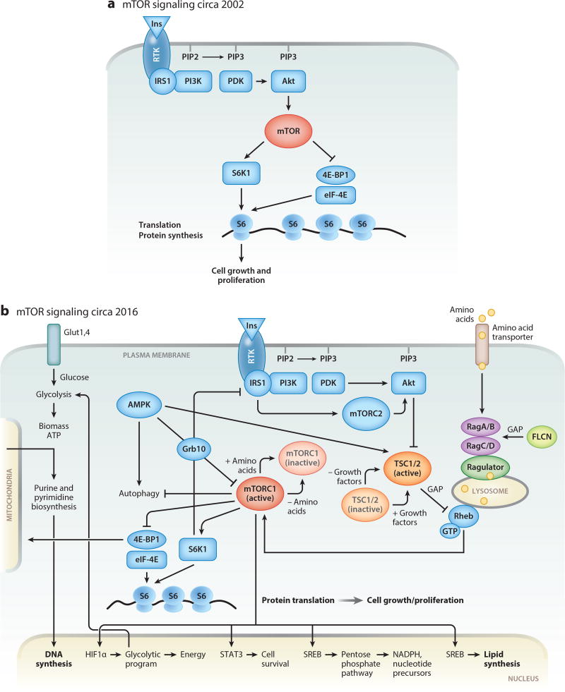
Lymphangioleiomyomatosis (LAM) is a rare, low-grade, metastasizing neoplasm that arises from an unknown source, spreads via the lymphatics, and targets the lungs. All pulmonary structures become infiltrated with benign-appearing spindle and epithelioid cells (LAM cells) that express smooth-muscle and melanocyte-lineage markers, harbor mTOR-activating mutations in tuberous sclerosis complex (TSC) genes, and recruit abundant stromal cells. Elaboration of lymphangiogenic growth factors and matrix remodeling enzymes by LAM cells enables their access to lymphatic channels and likely drives the cystic lung remodeling that often culminates in respiratory failure. Dysregulated cellular signaling results in a shift from oxidative phosphorylation to glycolysis as the preferred mode of energy generation, to allow for the accumulation of biomass required for cell growth and tolerance of nutrient-poor, anaerobic environments. Symptomatic LAM occurs almost exclusively in females after menarche, highlighting the central but as yet poorly understood role for sex-restricted anatomical structures and/or hormones in disease pathogenesis. LAM is an elegant model of malignancy because biallelic mutations at a single genetic locus confer all features that define cancer upon the LAM cell-metabolic reprogramming and proliferative signals that drive uncontrolled growth and inappropriate migration and invasion, the capacity to exploit the lymphatic circulation as a vehicle for metastasis and access to the lungs, and destruction of remote tissues. The direct benefit of the study of this rare disease has been the rapid identification of an effective FDA-approved therapy, and the collateral benefits have included elucidation of the pivotal roles of mTOR signaling in the regulation of cellular metabolism and the pathogenesis of cancer.
Keywords: lymphangiomyomatosis; perivascular epithelioid cell tumor (PECOMA); tuberous sclerosis; tumor suppressor syndrome.
Figures
References
-
- Johnson SR, Cordier JF, Lazor R, et al. European Respiratory Society guidelines for the diagnosis and management of lymphangioleiomyomatosis. Eur. Respir. J. 2010;35:14–26. - PubMed
Publication types
MeSH terms
Substances
Grants and funding
LinkOut - more resources
Full Text Sources
Other Literature Sources
Medical
Miscellaneous
